Copyright
©The Author(s) 2015.
World J Gastroenterol. Mar 7, 2015; 21(9): 2840-2847
Published online Mar 7, 2015. doi: 10.3748/wjg.v21.i9.2840
Published online Mar 7, 2015. doi: 10.3748/wjg.v21.i9.2840
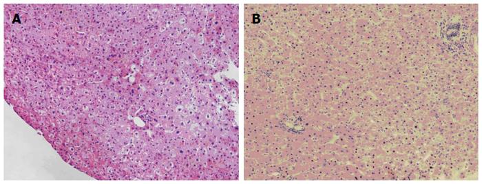
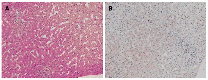
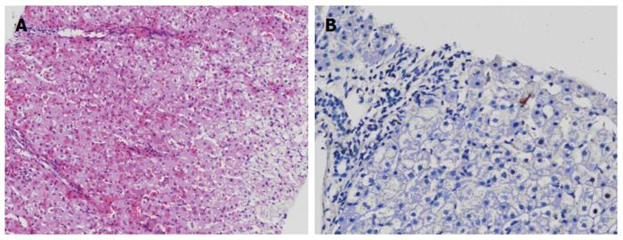
Figure 1 Liver biopsy prior to liver donation.
A: Normal liver structure (HE, × 100); B: No hepatocyte immunohistochemical staining for hepatitis B surface antigen (DAB, × 100).
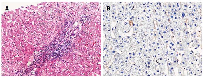
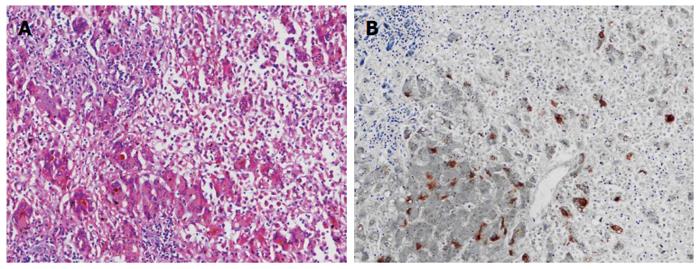
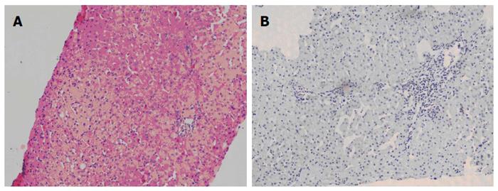
Figure 2 One month after liver transplantation.
A: Mild hepatitis with portal inflammation (HE, × 100); B: Immunohistochemical staining of cytoplasms for hepatitis B core antibody (DAB, × 100).
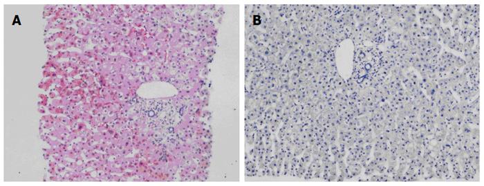
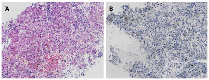
Figure 3 Three months after liver transplantation.
A: Return to almost normal liver structure (HE, × 40); B: No hepatocyte immunohistochemical staining for hepatitis B surface antigen (DAB, × 40).
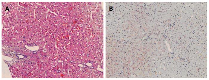
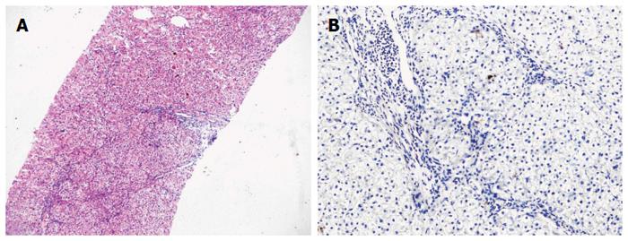
Figure 4 Six months after liver transplantation.
A: Almost normal liver structure with a little matrix deposition in portals (HE, × 100); B: No hepatocyte immunohistochemical staining for hepatitis B surface antigen (DAB, × 100).
Figure 5 One year after liver transplantation.
A: Almost normal liver structure (HE, × 100); B: No hepatocyte immunohistochemical staining for hepatitis B surface antigen (DAB, × 100).
Figure 6 Histology of recipient liver.
A: Acute severe hepatitis with massive necrosis (HE, × 100); B: Hepatocyte immunohistochemical staining for hepatitis B surface antigen (DAB, × 100).
Figure 7 Histology of recipient residual left liver one week later.
A: Acute hepatitis with less massive necrosis (HE, × 40); B: Hepatocyte immunohistochemical staining for hepatitis B surface antigen (DAB, × 40).
Figure 8 Histology of recipient residual left liver one month later.
A: Restored hepatic structure with several fine fibrous septa (HE, × 40); B: No hepatocyte immunohistochemical staining for hepatitis B surface antigen (DAB, × 100).
Figure 9 Histology of recipient residual left liver three months later.
A: Almost normal liver structure (HE, × 40); B: Hepatocyte immunohistochemical staining for hepatitis B surface antigen (DAB, × 40).
Figure 10 Histology of recipient residual left liver one year later.
A: Normal liver structure (HE, × 40); B: No hepatocyte immunohistochemical staining for hepatitis B surface antigen (DAB, × 40).
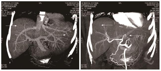
Figure 11 Computed tomography scan of fused liver one month after operation and 3-D reconstruction of hepatic vessels.
Donated right liver graft (solid arrows); Residual native left liver lobe (dotted arrows).
- Citation: Li CY, Lai W, Lu SC. Retrospective observation of therapeutic effects of adult auxiliary partial living donor liver transplantation on postpartum acute liver failure: A case report. World J Gastroenterol 2015; 21(9): 2840-2847
- URL: https://www.wjgnet.com/1007-9327/full/v21/i9/2840.htm
- DOI: https://dx.doi.org/10.3748/wjg.v21.i9.2840


