Copyright
©The Author(s) 2015.
World J Gastroenterol. Feb 28, 2015; 21(8): 2563-2567
Published online Feb 28, 2015. doi: 10.3748/wjg.v21.i8.2563
Published online Feb 28, 2015. doi: 10.3748/wjg.v21.i8.2563
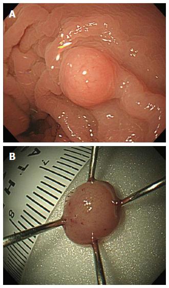
Figure 1 Colonoscopic finding.
A: Endoscopy revealed a hard, a yellow-white, solitary sessile mass (about 5 mm) at 10 cm from the anal verge of the rectum; B: Macroscopic view, after cap-endoscopic mucosal resection.
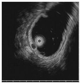
Figure 2 On endoscopic ultrasound: Well-demarcated, homogeneous hypoechoic solid lesion in the submucosal layer.
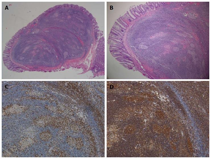
Figure 3 Histopathological findings.
A: Low-power view of the cap-endoscopic mucosal resection specimen. Hematoxylin and eosin (HE) staining, magnification × 20; B: HE staining, magnification × 40; C: Positive CD20 immunohistochemical staining, magnification × 100; D: Positive CD3 immunohistochemical staining, magnification × 100.
- Citation: Hong JB, Kim HW, Kang DH, Choi CW, Park SB, Kim DJ, Ji BH, Koh KW. Rectal tonsil: A case report and literature review. World J Gastroenterol 2015; 21(8): 2563-2567
- URL: https://www.wjgnet.com/1007-9327/full/v21/i8/2563.htm
- DOI: https://dx.doi.org/10.3748/wjg.v21.i8.2563


