Copyright
©The Author(s) 2015.
World J Gastroenterol. Feb 28, 2015; 21(8): 2558-2562
Published online Feb 28, 2015. doi: 10.3748/wjg.v21.i8.2558
Published online Feb 28, 2015. doi: 10.3748/wjg.v21.i8.2558
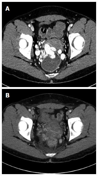
Figure 1 Computed tomography.
A: Abdominopelvic computed tomography. Huge rectal varices protrude into the rectum; B: Follow-up computed tomography at 5 d after variceal embolization. Obliteration of rectal varices is identified.
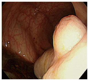
Figure 2 Sigmoidoscopic examination.
Huge rectal varices protrude into the rectum, with bleeding.
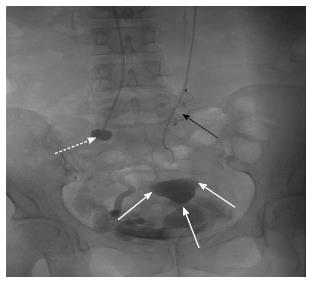
Figure 3 Angiography.
After applying an occlusion balloon (white dotted arrow) and vascular plug (black arrow), variceal embolization was successfully performed using a Gelfoam pledget (white arrow).
- Citation: Ahn SS, Kim EH, Kim MD, Lee WJ, Kim SU. Successful hemostasis of intractable rectal variceal bleeding using variceal embolization. World J Gastroenterol 2015; 21(8): 2558-2562
- URL: https://www.wjgnet.com/1007-9327/full/v21/i8/2558.htm
- DOI: https://dx.doi.org/10.3748/wjg.v21.i8.2558


