Copyright
©The Author(s) 2015.
World J Gastroenterol. Feb 28, 2015; 21(8): 2413-2418
Published online Feb 28, 2015. doi: 10.3748/wjg.v21.i8.2413
Published online Feb 28, 2015. doi: 10.3748/wjg.v21.i8.2413
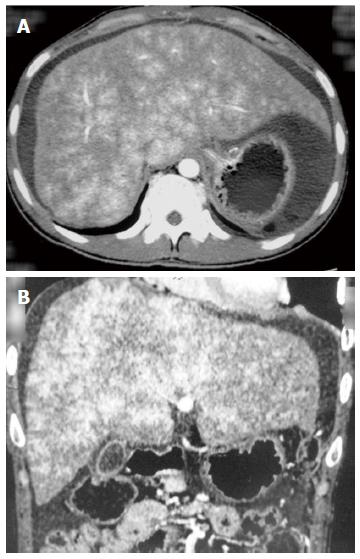
Figure 1 Abdominal computed tomography.
A and B: Enlargement, congestion and geographic change of the liver were noted.
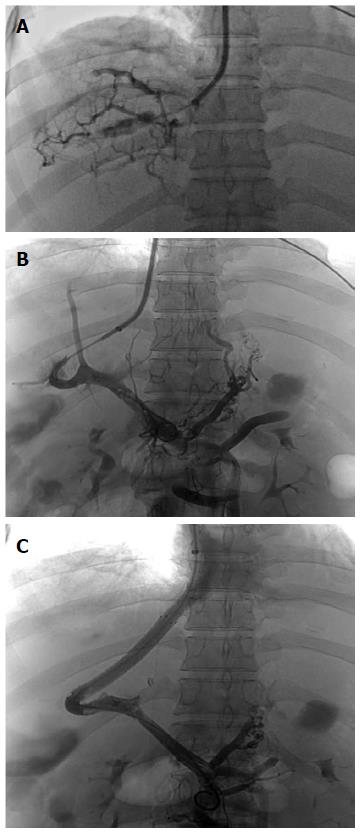
Figure 2 Transjugular intrahepatic portosystemic shunt.
A: Angiography revealed occluded hepatic venules; B: A guide wire was placed into the portal vein from the inferior vena cava and the varicose gastric coronary vein was noted by angiography of the portal vein; C: Transjugular intrahepatic portosystemic shunt was formed and the gastric coronary vein was embolized.
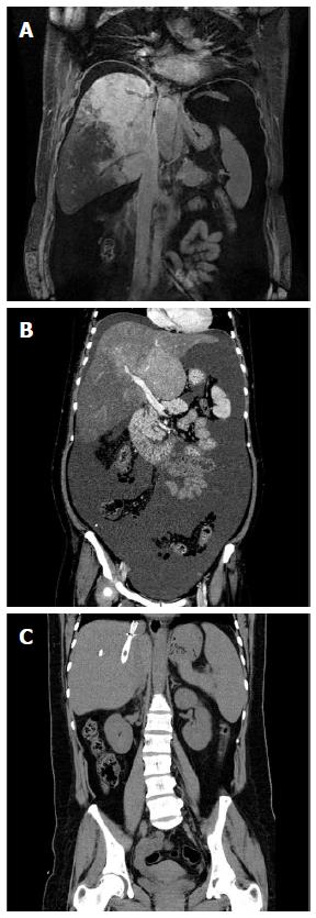
Figure 3 Coronal scans of magnetic resonance imaging of the portal vein and abdominal computed tomography of the same patient.
A and B: Magnetic resonance imaging of the portal vein and abdominal computed tomography revealed liver congestion and massive ascites before transjugular intrahepatic portosystemic shunt; C: Liver congestion and ascites disappeared 4 wk after the procedure.
- Citation: He FL, Wang L, Zhao HW, Fan ZH, Zhao MF, Dai S, Yue ZD, Liu FQ. Transjugular intrahepatic portosystemic shunt for severe jaundice in patients with acute Budd-Chiari syndrome. World J Gastroenterol 2015; 21(8): 2413-2418
- URL: https://www.wjgnet.com/1007-9327/full/v21/i8/2413.htm
- DOI: https://dx.doi.org/10.3748/wjg.v21.i8.2413


