Copyright
©The Author(s) 2015.
World J Gastroenterol. Feb 28, 2015; 21(8): 2323-2335
Published online Feb 28, 2015. doi: 10.3748/wjg.v21.i8.2323
Published online Feb 28, 2015. doi: 10.3748/wjg.v21.i8.2323
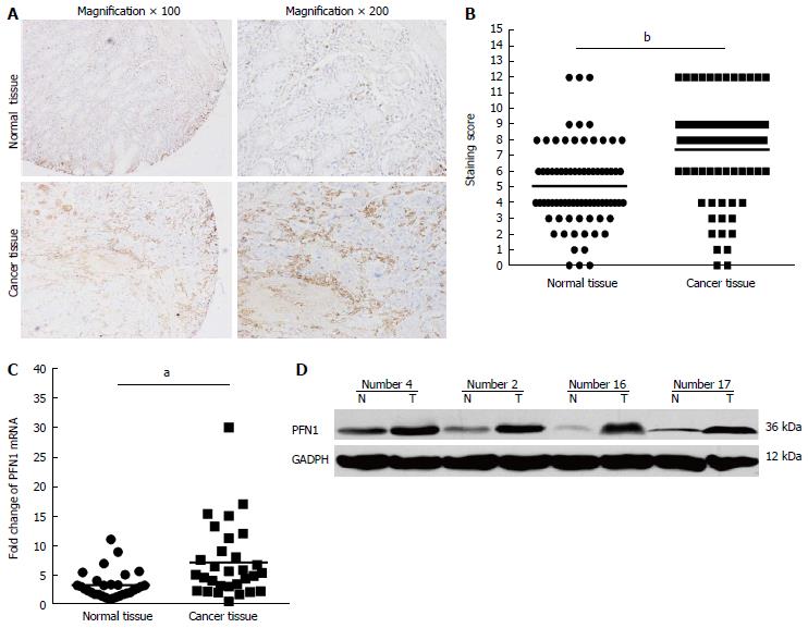
Figure 1 Analysis of profilin-1 expression in human gastric cancer and adjacent normal tissues.
A: Immunohistochemical staining for profilin-1 (PFN1) in human gastric cancer and adjacent normal tissues; B: The PFN1 staining score in human gastric cancer and adjacent normal tissues; C and D: Quantitative real-time polymerase chain reaction (C) and Western blot analyses (D) of PFN1 expression in another set of gastric cancer tumor tissues compared with paired adjacent normal tissues (n = 30) assessed. T: Tumor tissues; N: Adjacent normal tissues. aP < 0.05, bP < 0.01 vs paired adjacent normal tissues. GAPDH: Glyceraldehyde-3-phosphate dehydrogenase.
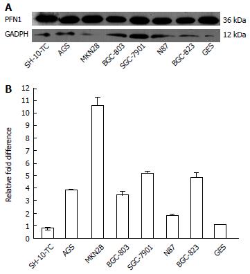
Figure 2 Analysis of profilin-1expression in human gastric cancer cell lines.
Western blot (A) and quantitative real-time polymerase chain reaction analyses (B) revealed the expression of profilin-1 (PFN1) in gastric cancer epithelial cell lines. GAPDH: Glyceraldehyde-3-phosphate dehydrogenase.
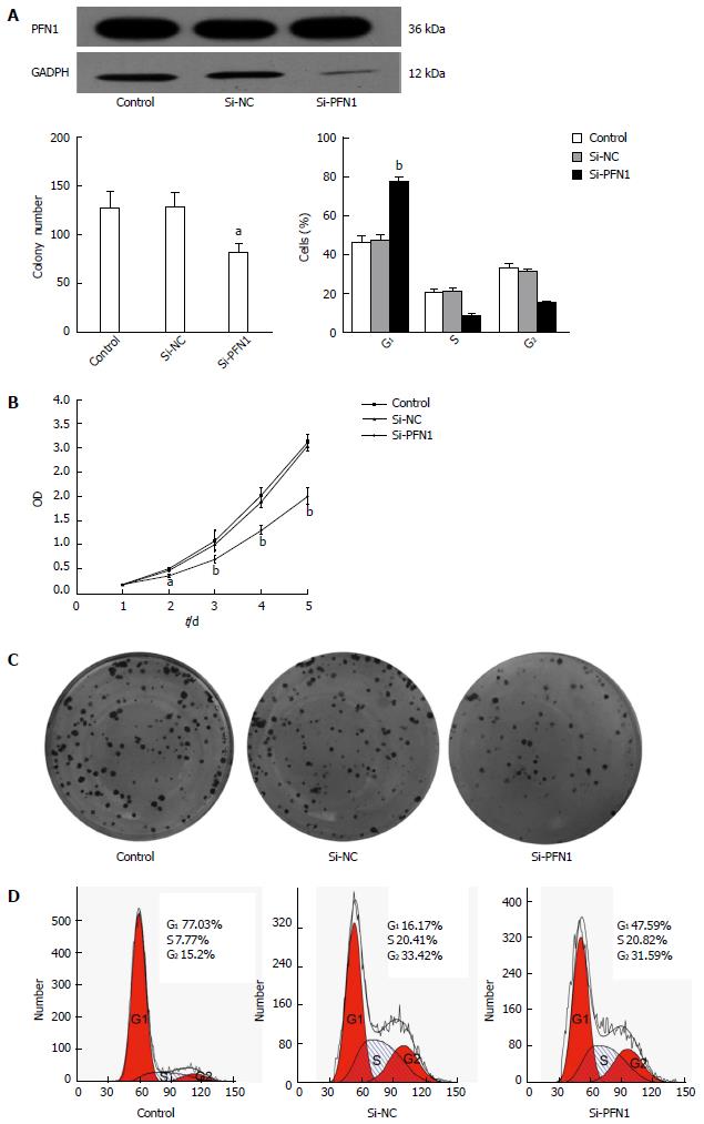
Figure 3 Profilin-1 knockdown inhibits tumor growth.
A: Expression of profilin-1 (PFN1) was assessed in the PFN1-transfected cells, the untreated control and the negative control (NC)-transfectants by Western blot; B: Cell proliferation levels were measured using the cell counting kit-8 on 1, 2, 3, 4 and 5 d. The optical density (A) represents the proliferative characteristics of the treated cells; C: PFN1 silencing resulted in significantly lower colony-forming efficiency; D: Cell-cycle distribution was analyzed by flow cytometry on SGC-7901 cells. The results obtained from three independent experiments are shown. aP < 0.05, bP < 0.01 vs other groups. GAPDH: Glyceraldehyde-3-phosphate dehydrogenase; Si: Silencing.
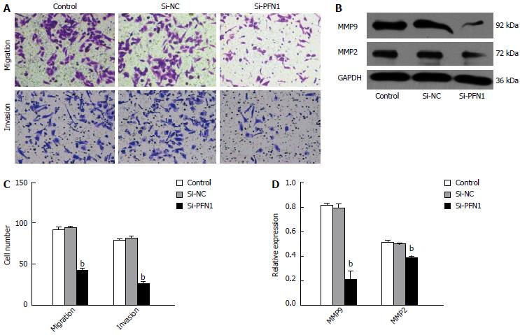
Figure 4 Effects of profilin-1 on gastric cancer cell migration in vitro.
A: The cell migration and invasion were assessed using transwell and invasion assays, respectively; B: The protein expression of matrix metalloproteinase (MMP)-2 and MMP9 was detected by Western blot. All results were obtained from three independent experiments. bP < 0.01 vs other groups. GAPDH: Glyceraldehyde-3-phosphate dehydrogenase; PFN1: Profilin-1; NC: Negative control; Si: Silencing.
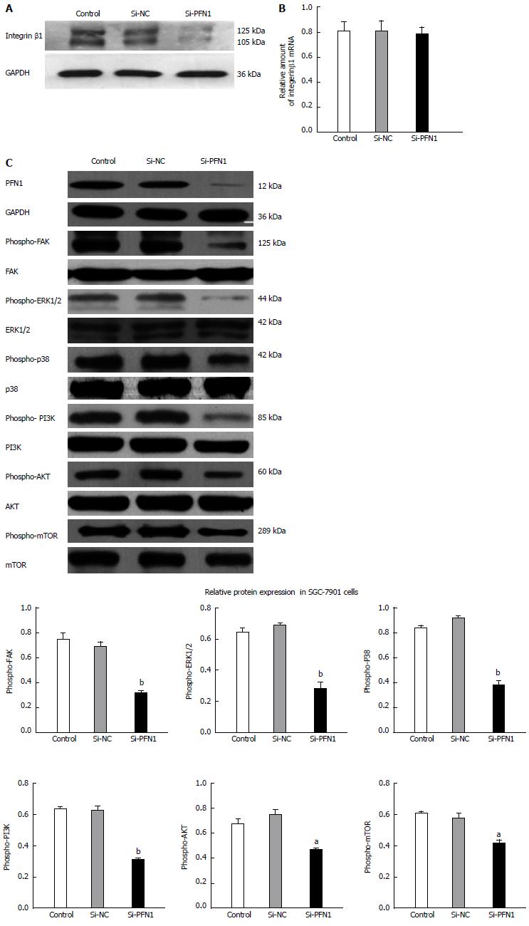
Figure 5 Profilin-1 silencing regulates the expression of integrin β1 and its downstream molecular targets.
A and B: Expression of integrin β1 protein and mRNA was detected by Western blot and quantitative real-time polymerase chain reaction analyses, respectively; C: The effect of profilin-1 (PFN1) knockdown on its downstream targets in the focal adhesion kinase (FAK) signaling pathway. The quantity of PFN1 was normalized against that of glyceraldehyde-3-phosphate dehydrogenase (GAPDH), and the phosphorylation of other proteins was normalized against the respective total protein. All results were obtained from three independent experiments. aP < 0.05, bP < 0.01 vs other groups. ERK: Extracellular-regulated kinase; mTOR: Mammalian target of rapamycin; PI3K: Phosphatidylinositol 3-kinase; GAPDH: Glyceraldehyde-3-phosphate dehydrogenase; NC: Negative control; Si: Silencing.
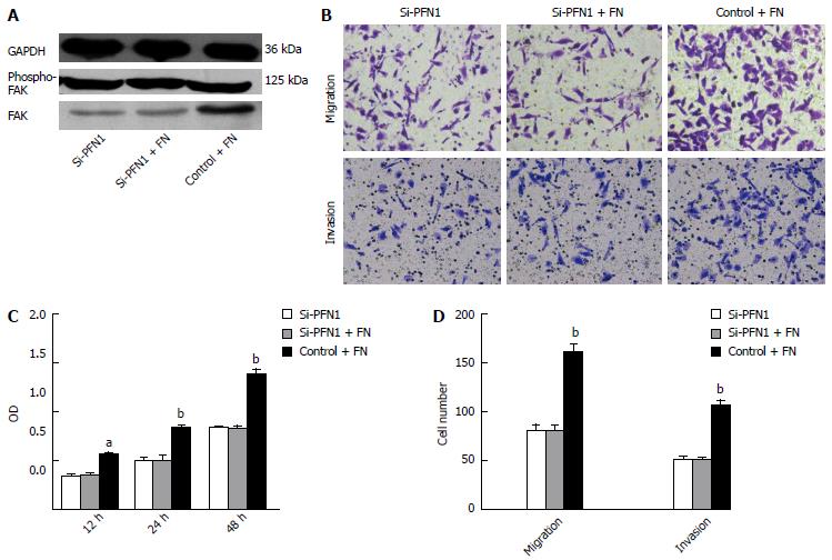
Figure 6 The effect of fibronectin is ameliorated after profilin-1 silencing.
A: SGC-7901 cells with and without profilin-1 (PFN1) silencing were seeded onto media with or without fibronectin (FN) (10 μg/mL) for 24 h. The expression of phosphorylated focal adhesion kinase (FAK) was evaluated by Western blot; B: SGC-7901 cells with and without PFN1 silencing were seeded on media with or without FN (10 μg/mL) at the indicated time points. Cell proliferation levels were measured using the cell counting kit-8 at 12, 24 and 48 h. The optical density (A) represents the proliferative characteristics of the treated cells; C: SGC-7901 cells with and without PFN1 silencing were suspended in media with or without FN (10 μg/mL) and seeded onto transwell plates. The cell migration was assessed 24 h later. For the invasion assays, the cells were seeded onto BD Matrigel invasion chambers. aP < 0.05, bP < 0.01 vs other groups. GAPDH: Glyceraldehyde-3-phosphate dehydrogenase; Si: Silencing.
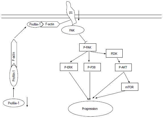
Figure 7 Schematic diagram of the mechanism underling the inhibition of gastric cancer progression through profilin-1 silencing.
Profilin-1 (PFN1) silencing decreases actin filament assembly and reduces the binding among PFN1, actin and integrin β1, which inhibits the focal adhesion kinase (FAK) activation through the down regulation of integrin β1 expression. FAK inhibition through profilin-1 silencing inhibits gastric cancer progression via mitogen-activated protein kinase and phosphatidylinositol 3-kinase (PI3K)/AKT signaling pathways. mTOR: Mammalian target of rapamycin.
-
Citation: Cheng YJ, Zhu ZX, Zhou JS, Hu ZQ, Zhang JP, Cai QP, Wang LH. Silencing profilin-1 inhibits gastric cancer progression
via integrin β1/focal adhesion kinase pathway modulation. World J Gastroenterol 2015; 21(8): 2323-2335 - URL: https://www.wjgnet.com/1007-9327/full/v21/i8/2323.htm
- DOI: https://dx.doi.org/10.3748/wjg.v21.i8.2323


