Copyright
©The Author(s) 2015.
World J Gastroenterol. Feb 28, 2015; 21(8): 2303-2314
Published online Feb 28, 2015. doi: 10.3748/wjg.v21.i8.2303
Published online Feb 28, 2015. doi: 10.3748/wjg.v21.i8.2303
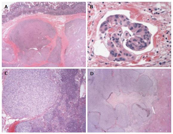
Figure 1 Histological features of succinate dehydrogenase-deficient gastrointestinal stromal tumors.
A: Multinodular architecture; B: Lymphovascular invasion; C: Lymph node metastasis; D: Liver metastasis with a multinodular/lobulated architecture similar to that seen in gastric primary SDH-deficient tumors. Data adapted from Barletta et al[50].
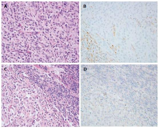
Figure 2 Morphological features of succinate dehydrogenase-deficient gastrointestinal stromal tumor cells.
A: Epithelioid gastrointestinal stromal tumor (GIST) cells (HE staining); B: Loss of expression of succinate dehydrogenase subunit B (SDHB, immunohistochemistry); C: Mixed epithelioid and spindle GIST cells (HE staining); D: Loss of expression of SDHB (immunohistochemistry). Data adapted from Wagner et al[25].
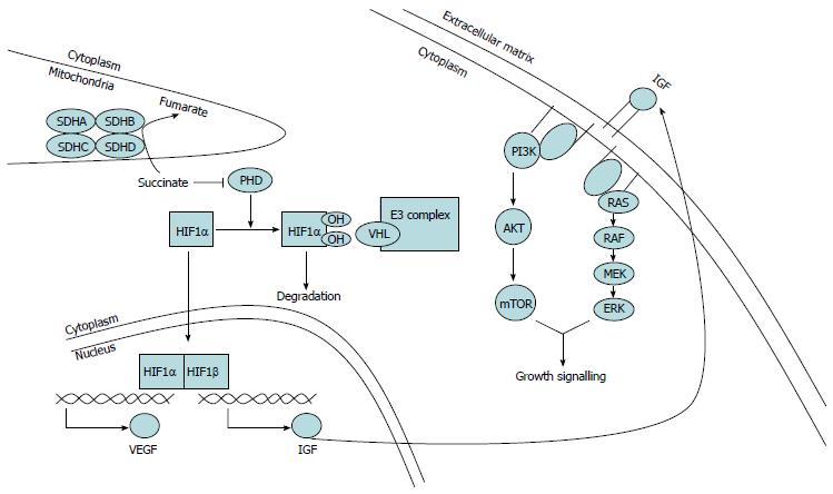
Figure 3 Role of succinate dehydrogenase deficiency in the tumorigenesis of succinate dehydrogenase-deficient gastrointestinal stromal tumors.
SDH complex dysfunction within mitochondria leads to increased levels of succinate, which in turn inhibits PHD activity. Non-hydroxylated HIF1α is resistant to VHL-mediated targeting for degradation and stimulates transcription of VEGF and IGF. SDH: Succinate dehydrogenase; HIF1α: Hypoxia-inducible factor α; PHD: Prolyl hydroxylase; E3 complex: Ubiquitin ligase complex; VHL: Von Hippel-Lindau; VEGF: Vascular endothelial growth factor; IGF: Insulin-like growth factor; PI3K: Phosphatidylinositol 3-kinase; mTOR: Mammalian target of rapamycin.
- Citation: Wang YM, Gu ML, Ji F. Succinate dehydrogenase-deficient gastrointestinal stromal tumors. World J Gastroenterol 2015; 21(8): 2303-2314
- URL: https://www.wjgnet.com/1007-9327/full/v21/i8/2303.htm
- DOI: https://dx.doi.org/10.3748/wjg.v21.i8.2303


