Copyright
©The Author(s) 2015.
World J Gastroenterol. Feb 7, 2015; 21(5): 1675-1679
Published online Feb 7, 2015. doi: 10.3748/wjg.v21.i5.1675
Published online Feb 7, 2015. doi: 10.3748/wjg.v21.i5.1675
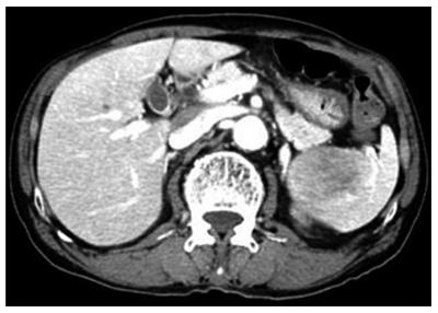
Figure 1 Computed tomography of the abdomen and pelvis shows a 5.
7 cm × 4.5 cm sized solid mass in the spleen.
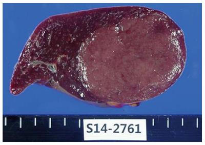
Figure 2 A specimen obtained during splenectomy.
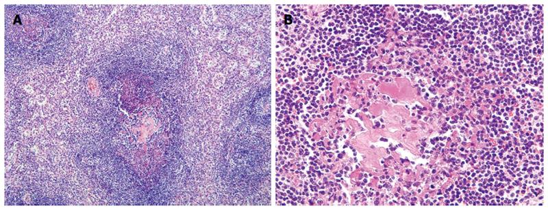
Figure 3 Castleman’s disease.
A: In a low-power field (magnification × 100), enlarged follicles with atrophic centers and pale pink vessels rich in interfollicular stroma are observed; B: In a high-power field (magnification × 400), hyaline deposition in the germinal center and expanded mantle zones of concentric small lymphocytes are observed.
- Citation: Lee HJ, Jeon HJ, Park SG, Park CY. Castleman’s disease of the spleen. World J Gastroenterol 2015; 21(5): 1675-1679
- URL: https://www.wjgnet.com/1007-9327/full/v21/i5/1675.htm
- DOI: https://dx.doi.org/10.3748/wjg.v21.i5.1675


