Copyright
©The Author(s) 2015.
World J Gastroenterol. Feb 7, 2015; 21(5): 1670-1674
Published online Feb 7, 2015. doi: 10.3748/wjg.v21.i5.1670
Published online Feb 7, 2015. doi: 10.3748/wjg.v21.i5.1670
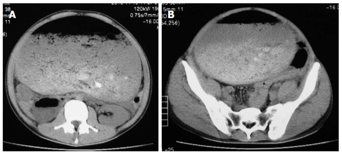
Figure 1 Computed tomography on initial admission.
A. Upper abdomen B. Pelvis.
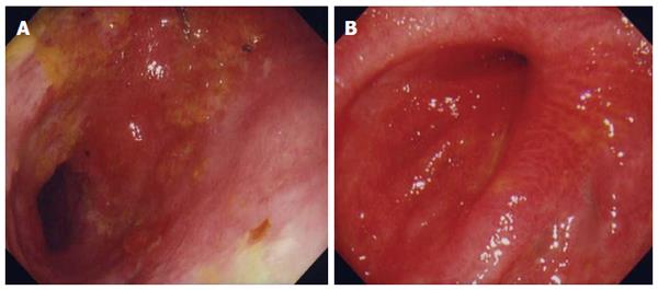
Figure 2 Gastrointestinal endoscopy revealed pyloric stenosis.
A: One month after admission, gastrointestinal endoscopy revealed mucosal deciduation at the pyloric antrum, but there were no findings of pyloric stenosis; B: One year after discharge, gastrointestinal endoscopy revealed progressive pyloric stenosis.
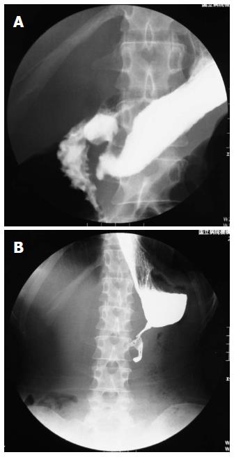
Figure 3 Gastrointestinal series showed pyloric stenosis.
A: Two months after admission, an upper gastrointestinal series showed no leakage and a good passage of contrast medium; B: One year after discharge, a follow-up gastrointestinal series showed advanced pyloric stenosis.
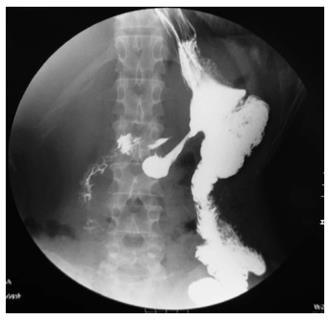
Figure 4 A postoperative gastrointestinal series showed improvement in the contrast medium passage through the stomach and intestine.
- Citation: Kimura A, Masuda N, Haga N, Ito T, Otsuka K, Takita J, Satomura H, Kumakura Y, Kato H, Kuwano H. Gastrojejunostomy for pyloric stenosis after acute gastric dilatation due to overeating. World J Gastroenterol 2015; 21(5): 1670-1674
- URL: https://www.wjgnet.com/1007-9327/full/v21/i5/1670.htm
- DOI: https://dx.doi.org/10.3748/wjg.v21.i5.1670


