Copyright
©The Author(s) 2015.
World J Gastroenterol. Feb 7, 2015; 21(5): 1588-1594
Published online Feb 7, 2015. doi: 10.3748/wjg.v21.i5.1588
Published online Feb 7, 2015. doi: 10.3748/wjg.v21.i5.1588
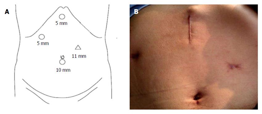
Figure 1 Placement of trocars (A) and postoperative image (B).
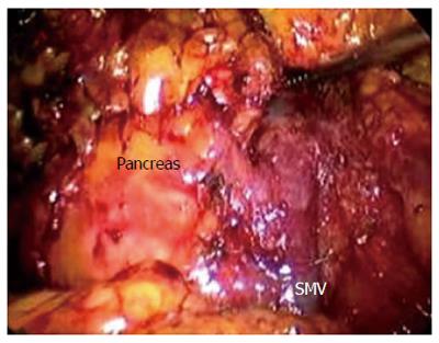
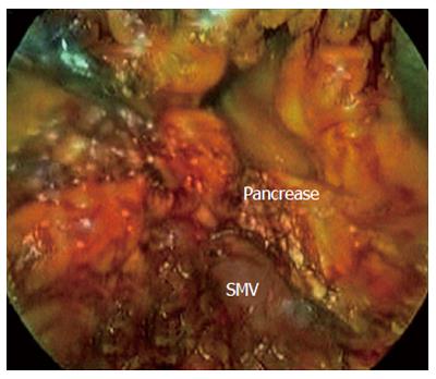
Figure 2 Freeing the gastrocolic ligament exposes the inferior border of the pancreas and superior mesenteric vein.
SMV: Superior mesenteric vein.
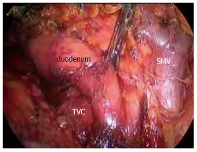
Figure 3 Mobilization of the duodenum and the proximal jejunum from right to left under the superior mesenteric vein (mobilization of the duodenojejunal flexure from the retroperitoneum).
SMV: Superior mesenteric vein;
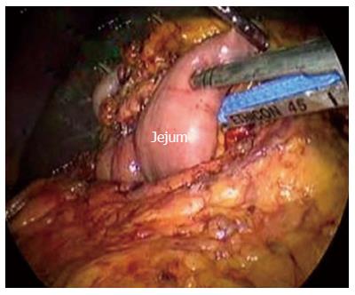
Figure 4 Transection of the jejunum.
Figure 5 Transection of the pancreas.
Pancreatic neck parenchyma is divided with ultrasonic shears. SMV: Superior mesenteric vein.
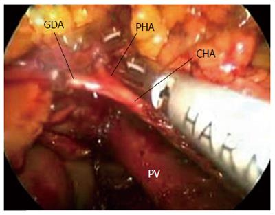
Figure 6 Common hepatic artery and the gastroduodenal and right gastric arteries are identified and isolated from posterior to anterior.
CHA: Common hepatic artery; PV: Portal vein.
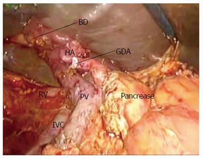
Figure 7 After the pancreatic-duodenum removal.
PV: Portal vein;
-
Citation: Liu Z, Yu MC, Zhao R, Liu YF, Zeng JP, Wang XQ, Tan JW. Laparoscopic pancreaticoduodenectomy
via a reverse-''V'' approach with four ports: Initial experience and perioperative outcomes. World J Gastroenterol 2015; 21(5): 1588-1594 - URL: https://www.wjgnet.com/1007-9327/full/v21/i5/1588.htm
- DOI: https://dx.doi.org/10.3748/wjg.v21.i5.1588


