Copyright
©The Author(s) 2015.
World J Gastroenterol. Feb 7, 2015; 21(5): 1468-1478
Published online Feb 7, 2015. doi: 10.3748/wjg.v21.i5.1468
Published online Feb 7, 2015. doi: 10.3748/wjg.v21.i5.1468
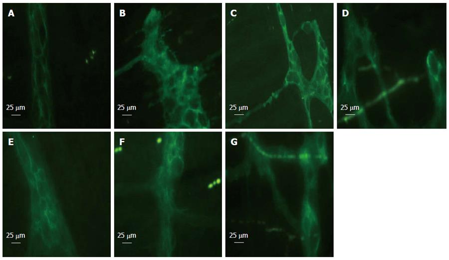
Figure 1 Glial fibrillary acidic protein immunofluorescent staining at 6 h after blood loss.
Enteric glial cells (EGCs) were distorted following hemorrhage and showed morphological abnormalities. EGCs presented as normal in the sham group. Electroacupuncture (EA) ST36 attenuated the morphological changes. After hemorrhagic shock (HS), the fluorescent intensity was enhanced. EA ST36 showed more intense fluorescence of glial fibrillary acidic protein (GFAP) staining, whereas fluorescence after VGX or injection of α-bungarotoxin (BGT) was weak. All images were taken at magnification × 400; white bar = 25 μm (n = 3-6 rats per group at 6 h after blood loss). A: Sham group; B: HS group; C: EA group; D: VGX/EA group; E: VGX group; F: α-BGT/EA; G: α-BGT group.
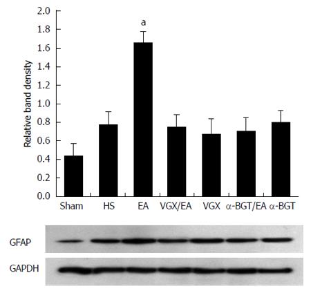
Figure 2 Glial fibrillary acidic protein expression at 6 h after blood loss.
Intestinal extracts were obtained from rats at 6 h after blood loss for measurement of glial fibrillary acidic protein expression using western blotting. Representative blots for glial fibrillary acidic protein (GFAP) are shown, with the corresponding GAPDH loading control to demonstrate equal protein load in all lanes. The GFAP level was lower in the sham group. EA ST36 increased GFAP expression (aP < 0.05 vs HS group). Significantly weak GFAP expression was seen in all the other injury groups (n = 3-6 rats per group at 6 h after blood loss). α-BGT: α-bungarotoxin; VGX: Vagotomy; EA: Electroacupuncture; HS: Hemorrhagic shock.
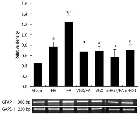
Figure 3 Electroacupuncture ST 36 alters expression of intestinal glial fibrillary acidic protein.
RT-PCR was performed on intestinal extracts obtained 6 h following injury. EA ST 36 resulted in augmented intestinal glial fibrillary acidic protein (GFAP) mRNA expression in the EA group compared with the HS group. Abdominal vagotomy (VGX) or injection of α-bungarotoxin (BGT) abrogated the effect of EA ST36 to increase intestinal GFAP mRNA expression. aP < 0.05 vs sham group, cP < 0.05 vs HS group. EA: Electroacupuncture; HS: Hemorrhagic shock.
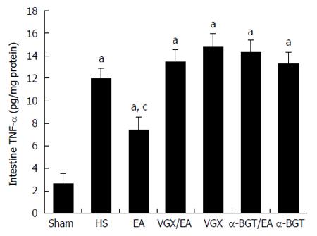
Figure 4 Tumor necrosis factor-α levels in intestine at 6 h after blood loss.
The intestine was obtained at 6 h after blood loss. Data are expressed as mean ± SD (n = 3-6 rats at every time point per group). aP < 0.05 vs sham group; cP < 0.05 vs hemorrhagic shock (HS) group. α-BGT: α-bungarotoxin; VGX: Vagotomy; EA: Electroacupuncture; TNF-α: Tumor necrosis factor-α.
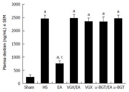
Figure 5 Intestinal permeability to 4-kDa fluorescein isothiocyanate-dextran at 6 h after blood loss.
EA ST36 protected the intestine from an increase in permeability after hemorrhagic shock (HS), whereas EA ST36 after vagotomy (VGX) or injection of α-bungarotoxin (BGT) eliminated such protection. aP < 0.05 vs sham group, cP < 0.05 vs HS group (n = 3-6 animals per group at 6 h after blood loss). EA: Electroacupuncture.
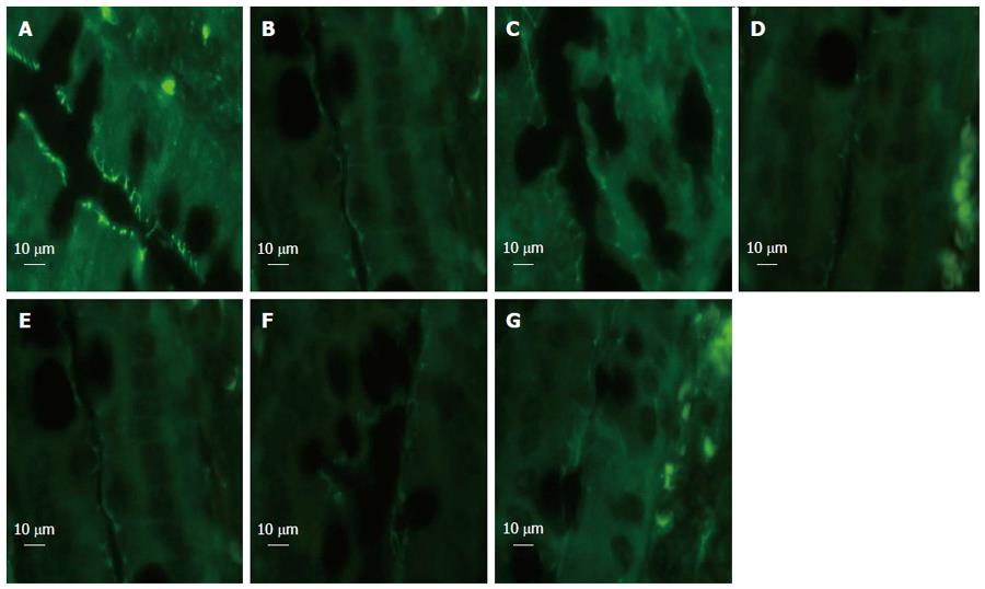
Figure 6 Intestinal zona occludens-1 immunofluorescent staining at 6 h after blood loss.
In the sham group, zona occludens (ZO)-1 presented with strong fluorescent intensity. Rats in the hemorrhagic shock (HS) group showed low fluorescent intensity at the cell periphery after HS, and electroacupuncture (EA) ST36 showed preservation of the robust structure of ZO-1 staining, whereas EA ST36 after vagotomy (VGX) or injection of α-bungarotoxin (BGT) eliminated such protection. All images were taken at magnification × 1000 magnification; white bar = 10 μm. (n = 3-6 rats per group at 6 h after blood loss, section size = 2 μm). A: Sham group; B: HS group; C: EA group; D: VGX/EA group; E: VGX group; F: α-BGT/EA; G: α-BGT group.
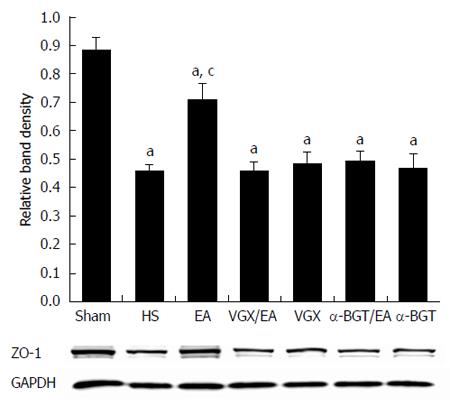
Figure 7 Intestinal zona occludens-1 protein expression at 6 h after blood loss.
Intestinal extracts were obtained from rats at 6 h after blood loss to measure zona occludens (ZO)-1 protein expression using western blotting. Representative blots for ZO-1 protein are shown with corresponding GAPDH loading control to demonstrate equal protein load in all lanes. Electroacupuncture (EA) ST36 resulted in preservation of protein expression. Significant reduction in ZO-1 expression was seen in all the other groups. aP < 0.05 vs sham group; cP < 0.05 vs hemorrhagic shock (HS) group. (n = 3-6 rats per group at 6 h after blood loss). α-BGT: α-bungarotoxin; VGX: Vagotomy.
- Citation: Hu S, Zhao ZK, Liu R, Wang HB, Gu CY, Luo HM, Wang H, Du MH, Lv Y, Shi X. Electroacupuncture activates enteric glial cells and protects the gut barrier in hemorrhaged rats. World J Gastroenterol 2015; 21(5): 1468-1478
- URL: https://www.wjgnet.com/1007-9327/full/v21/i5/1468.htm
- DOI: https://dx.doi.org/10.3748/wjg.v21.i5.1468


