Copyright
©The Author(s) 2015.
World J Gastroenterol. Dec 21, 2015; 21(47): 13302-13308
Published online Dec 21, 2015. doi: 10.3748/wjg.v21.i47.13302
Published online Dec 21, 2015. doi: 10.3748/wjg.v21.i47.13302
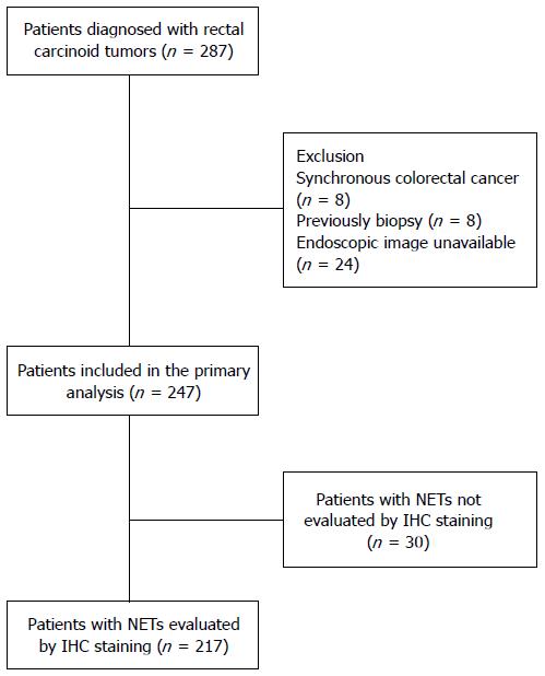
Figure 1 Study flow chart.
NETs: Neuroendocrine tumors; IHC: immunohistochemistry.
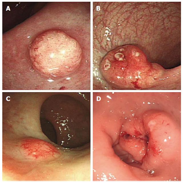
Figure 2 Endoscopic findings of atypical carcinoids.
A: Semipedunculated type with hyperemia; B: Semipedunculated type with erosion and hyperemia; C: Sessile type with hyperemia; D: An ulcerofungating types mimicking rectal cancer.
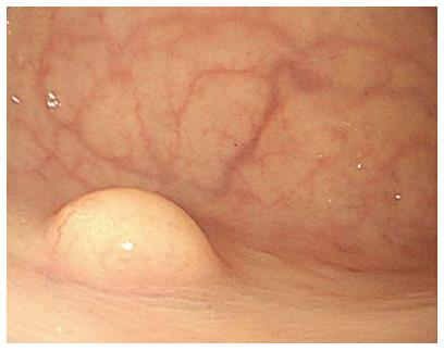
Figure 3 Endoscopic image of a typical carcinoid, which was a sessile tumor with a yellow, smooth surface.
- Citation: Hyun JH, Lee SD, Youk EG, Lee JB, Lee EJ, Chang HJ, Sohn DK. Clinical impact of atypical endoscopic features in rectal neuroendocrine tumors. World J Gastroenterol 2015; 21(47): 13302-13308
- URL: https://www.wjgnet.com/1007-9327/full/v21/i47/13302.htm
- DOI: https://dx.doi.org/10.3748/wjg.v21.i47.13302


