Copyright
©The Author(s) 2015.
World J Gastroenterol. Dec 21, 2015; 21(47): 13212-13224
Published online Dec 21, 2015. doi: 10.3748/wjg.v21.i47.13212
Published online Dec 21, 2015. doi: 10.3748/wjg.v21.i47.13212
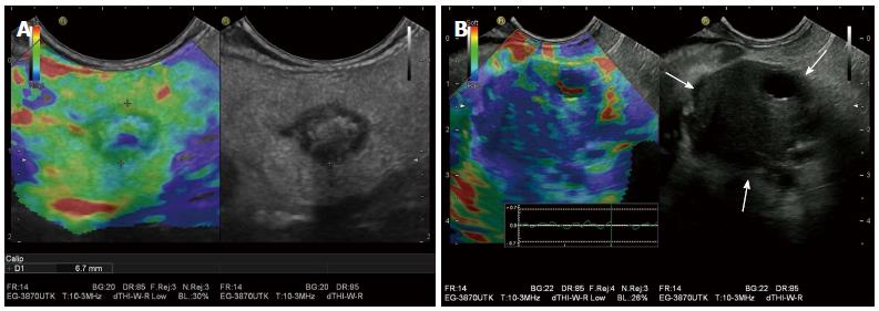
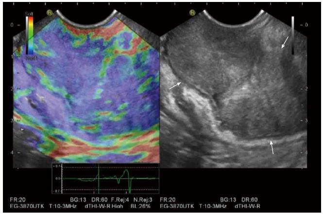
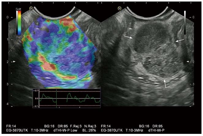
Figure 1 Benign and malignant pancreatic masses on endoscopic ultrasound elastography.
A: A pancreatic teratoma is shown as heterogeneous soft (green) pattern (left: EUS elastography image; right: B-mode image); B: A pancreatic ductal adenocarcinoma appears stiffer (blue) than the adjacent normal pancreatic parenchyma, probably due to the presence of fibrosis and marked desmoplasia.
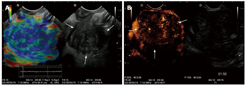
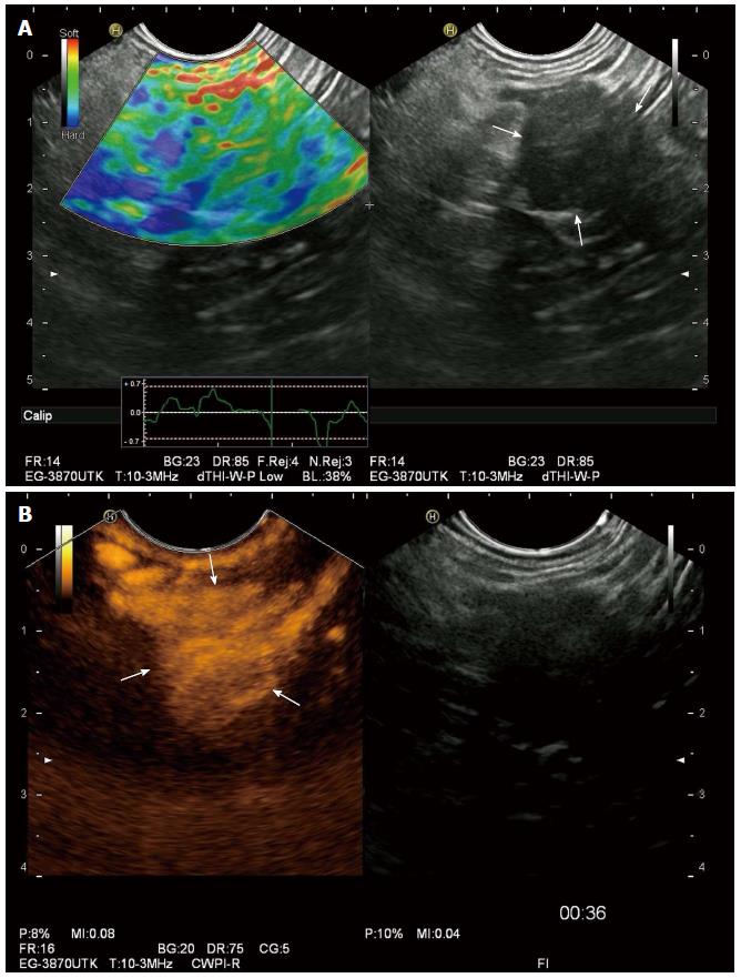
Figure 2 Combination of elastography and contrast-enhanced ultrasound in endoscopic ultrasound.
A: A pancreatic ductal adenocarcinoma is demonstrated as a heterogeneous hard (blue) pattern; B: On contrast-enhanced endoscopic ultrasound, pancreatic ductal adenocarcinoma is hypo-enhancing.
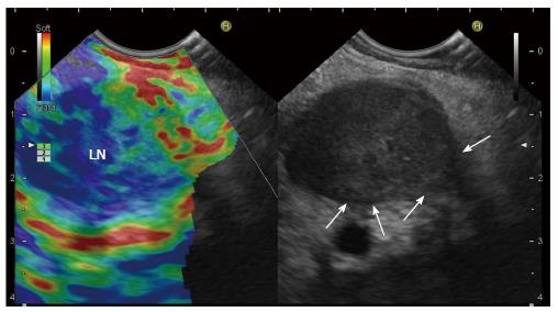
Figure 3 Malignant Lymph nodes on endoscopic ultrasound elastography.
A malignant lymph node is revealed as predominantly hard (blue). Arrows indicate infiltration. LN: Lymph node.
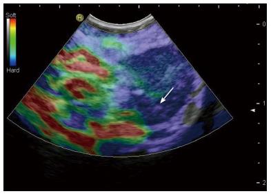
Figure 4 Endoscopic ultrasound elastography adds information to the B-mode evaluation of lymph nodes and can better guide a fine needle aspiration procedure by identifying stiffer (blue in the image) and thus, most suspicious regions for malignant infiltration.
Arrow indicates needle tip.
Figure 5 Gastric carcinoma is shown as heterogeneously stiff pattern (blue) on endoscopic ultrasound elastography.
Figure 6 Gastrointestinal stromal tumor is revealed heterogeneous hard (blue) pattern on endoscopic ultrasound elastography.
Figure 7 Benign adrenal gland tumor is typically soft (green) on endoscopic ultrasound elastography (A) and hyper-enhancing on contrast-enhanced endoscopic ultrasound (B).
- Citation: Cui XW, Chang JM, Kan QC, Chiorean L, Ignee A, Dietrich CF. Endoscopic ultrasound elastography: Current status and future perspectives. World J Gastroenterol 2015; 21(47): 13212-13224
- URL: https://www.wjgnet.com/1007-9327/full/v21/i47/13212.htm
- DOI: https://dx.doi.org/10.3748/wjg.v21.i47.13212


