Copyright
©The Author(s) 2015.
World J Gastroenterol. Dec 14, 2015; 21(46): 12996-13003
Published online Dec 14, 2015. doi: 10.3748/wjg.v21.i46.12996
Published online Dec 14, 2015. doi: 10.3748/wjg.v21.i46.12996
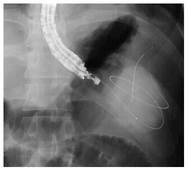
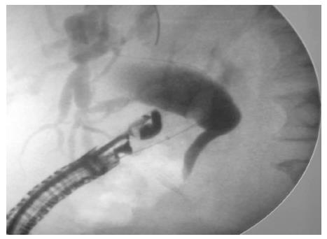
Figure 1 Fluoroscopic image showing a 0.
035-inch guidewire coiling within a pancreatic pseudocyst. Courtesy of Prof. Lee[18] with permission.
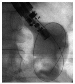
Figure 2 Fluoroscopic image showing dilation of the fistula tract with a balloon dilator.
Courtesy of Prof. Kahaleh[16] with permission.
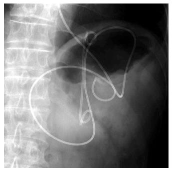
Figure 3 Fluoroscopic image showing placement of a 10-Fr double pig-tail plastic stent and a nasocystic catheter into a pancreatic pseudocyst.
Courtesy of Prof. Itoi[37] with permission.
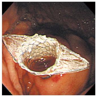
Figure 4 Endoscopic image of placement of a fully-covered metal stent into a pancreatic pseudocyst.
Courtesy of Prof. Lee[18] with permission.
Figure 5 Flouroscopic image of the choledochoduodenal tract dilation with a balloon dilator.
Courtesy of Prof. Altonbary[33] with permission.
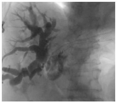
Figure 6 Flouroscopic image of placement of a metal stent into the hepatic biliary system.
Courtesy of Prof. Kahaleh[31] with permission.
- Citation: Meng FS, Zhang ZH, Ji F. Therapeutic role of endoscopic ultrasound in pancreaticobiliary disease: A comprehensive review. World J Gastroenterol 2015; 21(46): 12996-13003
- URL: https://www.wjgnet.com/1007-9327/full/v21/i46/12996.htm
- DOI: https://dx.doi.org/10.3748/wjg.v21.i46.12996


