Copyright
©The Author(s) 2015.
World J Gastroenterol. Dec 7, 2015; 21(45): 12970-12975
Published online Dec 7, 2015. doi: 10.3748/wjg.v21.i45.12970
Published online Dec 7, 2015. doi: 10.3748/wjg.v21.i45.12970
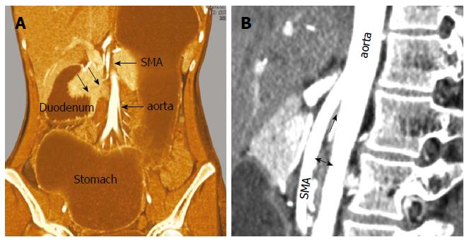
Figure 1 Abdominal computed tomography.
A: A distended stomach and proximal duodenum were shown with caliber change at the third portion (arrows), between superior mesenteric artery (SMA) and aorta; B: On the sagittal view, the aorto-mesenteric angle was 10° (arrow) and the aorto-mesenteric distance (two-head arrow) was 5.5 mm.
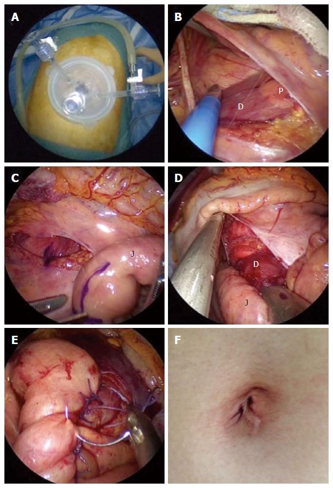
Figure 2 Intraoperative findings.
A: Three 5-mm trocars were inserted through the umbilical incision; B: Identification of the anterior wall of the second portion of the duodenum and pancreas; C: Duodenum and proximal jejunum (25 cm from Treitz ligament) were marked with crystal violet at the planned anastomosis site; D: 45-mm linear stapler was inserted to make a side-to-side duodenojejunostomy with duodenum and jejunum; E: The common entry hole was sutured by hand; F: The umbilical incision became virtually scarless 3 mo after the operation. D: Duodenum; J: Jejunum; P: Pancreas.
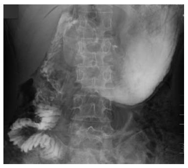
Figure 3 Contrast study on post-operative day 3 showed smooth fluid passage through the duodenojejunostomy.
- Citation: Yao SY, Mikami R, Mikami S. Minimally invasive surgery for superior mesenteric artery syndrome: A case report. World J Gastroenterol 2015; 21(45): 12970-12975
- URL: https://www.wjgnet.com/1007-9327/full/v21/i45/12970.htm
- DOI: https://dx.doi.org/10.3748/wjg.v21.i45.12970


