Copyright
©The Author(s) 2015.
World J Gastroenterol. Nov 28, 2015; 21(44): 12644-12652
Published online Nov 28, 2015. doi: 10.3748/wjg.v21.i44.12644
Published online Nov 28, 2015. doi: 10.3748/wjg.v21.i44.12644
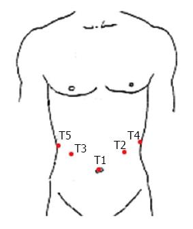
Figure 1 Placement of the 5 ports.
T1: 12 mm telescope trocar in the navel; T2: 12 mm trocar along the left midclavear lines; T3: 12-mm trocar along the right midclavear lines; T4: 5 mm trocar along the left anterior axillary line; T5: 5 mm trocar along the right anterior axillary line.
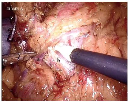
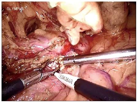
Figure 2 Skeletonized gastrocolic trunk and its branches.
a: Anterior superior pancreaticoduodenal vein; b: Right colic vein.
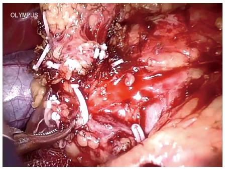
Figure 3 Resection of the branches of the gastrocolic trunk to expose the whole pancreatic head.
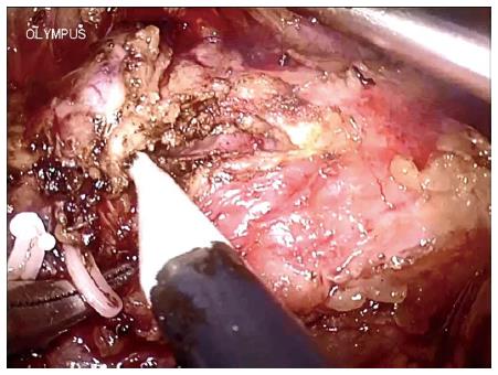
Figure 4 The pancreatic duct was opened longitudinally using the electrosurgical hook.
Figure 5 Large ductal stones are extracted while opening the duct distally and proximally.
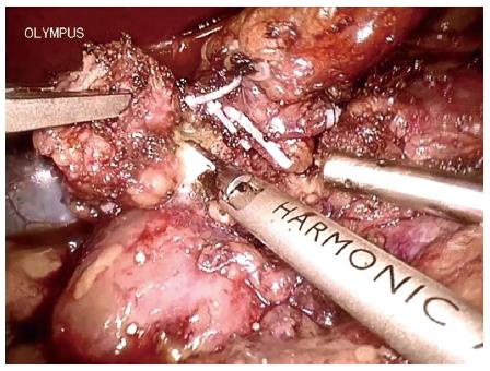
Figure 6 The parenchyma of the pancreatic head is excavated using a harmonic scalpel.
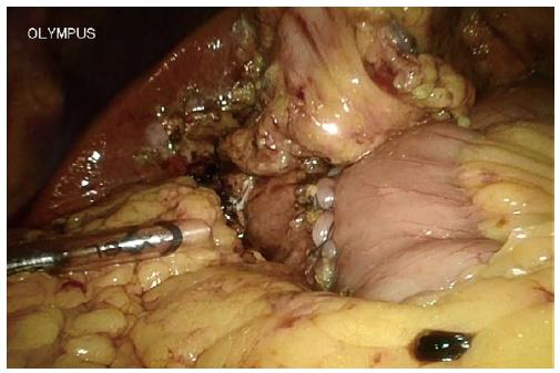
Figure 7 One layer side-to-side pancreaticojejunostomy completed with interrupted sutures.
- Citation: Tan CL, Zhang H, Li KZ. Single center experience in selecting the laparoscopic Frey procedure for chronic pancreatitis. World J Gastroenterol 2015; 21(44): 12644-12652
- URL: https://www.wjgnet.com/1007-9327/full/v21/i44/12644.htm
- DOI: https://dx.doi.org/10.3748/wjg.v21.i44.12644


