Copyright
©The Author(s) 2015.
World J Gastroenterol. Nov 21, 2015; 21(43): 12482-12497
Published online Nov 21, 2015. doi: 10.3748/wjg.v21.i43.12482
Published online Nov 21, 2015. doi: 10.3748/wjg.v21.i43.12482
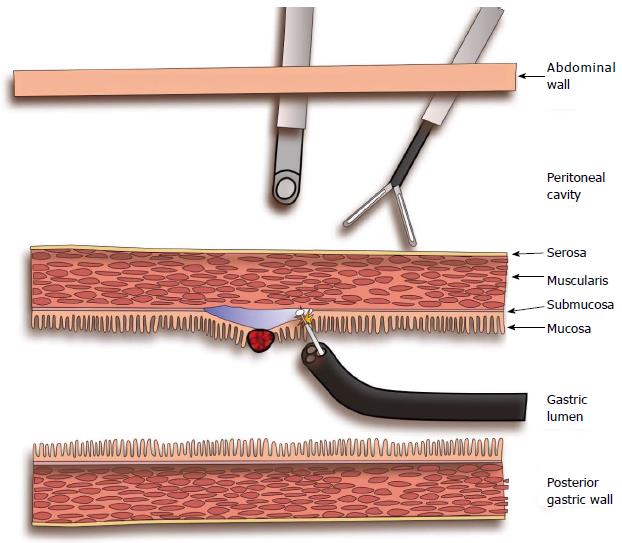
Figure 1 Laparoscopy-assisted endoscopic resection.
The endoscopist is performing an endoscopic submucosal dissection while the laparoscopic team facilitates the exposure by external manipulations.
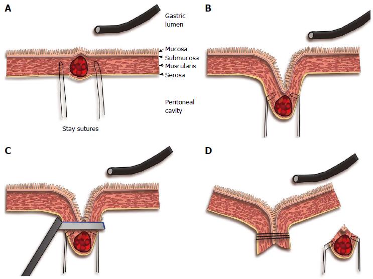
Figure 2 Endoscope-assisted laparoscopic wedge resection.
A: The tumor is located by endoscopy and laparoscopy; two laparoscopic stay sutures are placed; B: Traction by the stay sutures creates tenting of the gastric wall; C: An endoscopic stapler is applied at the normal gastric wall distally to the tumor under endoscopic control; D: Full thickness resection with everted stapling of the gastric wall.
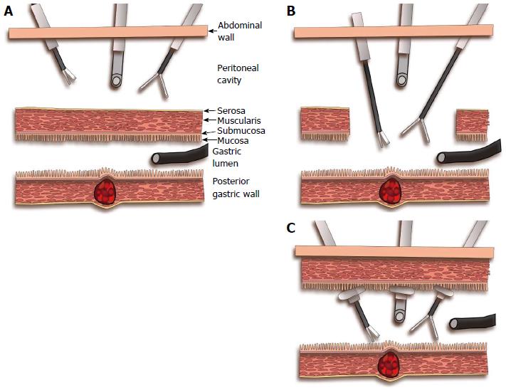
Figure 3 Endoscope-assisted laparoscopic intragastric and transgastric resection technique.
A: Localization of the tumor at the posterior gastric wall by endoscopy and the position of the anterior gastric wall overlying the tumor is shown to the laparoscopic team by transillumination; B: Transgastric resection: the anterior gastric wall is sectioned (gastrotomy) to give laparoscopic access to the gastric lumen and the tumor; C: Intragastric resection: the laparoscopic ports are inserted through the abdominal wall and through the gastric wall inside the gastric lumen; the gastric wall is held in contact with the abdominal wall by means of the balloon tipped laparoscopic ports.
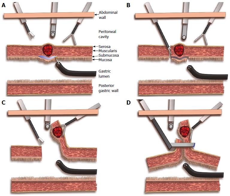
Figure 4 Laparoscopic endoscopic combined surgery.
A: Submucosal injection and section of the mucosa around the tumor by the endoscopic team; B: After perforation of the gastric wall by the endoscopic team, the laparoscopic team makes a full thickness incision following the path of the endoscopic resection; C: Eversion of the tumor towards the peritoneal cavity and laparoscopic resection around the 2/3 of the gastric wall around it; D: Resection of the tumor and closure of the gastric wall by application of a laparoscopic stapler.
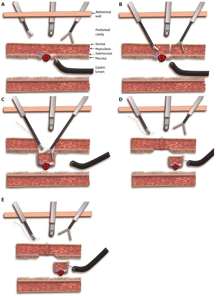
Figure 5 Non-exposed endoscopic wall-inversion surgery.
A: Endoscopic submucosal injection around the tumor; B: Laparoscopic seromuscular dissection around the tumor down to the stained submucosal plane; C: Retraction of the tumor with its overlying normal gastric wall towards the gastric lumen, and closure of the gastric wall by laparoscopic seromuscular extramucosal suturing; D: The gastric wall opening is completely closed and the tumor is invaginated inside the gastric lumen; endoscopic mucosal resection around the tumor with a needle knife. E: Endoscopic extraction of the tumor, and the gastric mucosa is approximated with clips.
- Citation: Ntourakis D, Mavrogenis G. Cooperative laparoscopic endoscopic and hybrid laparoscopic surgery for upper gastrointestinal tumors: Current status. World J Gastroenterol 2015; 21(43): 12482-12497
- URL: https://www.wjgnet.com/1007-9327/full/v21/i43/12482.htm
- DOI: https://dx.doi.org/10.3748/wjg.v21.i43.12482


