Copyright
©The Author(s) 2015.
World J Gastroenterol. Nov 7, 2015; 21(41): 11854-11861
Published online Nov 7, 2015. doi: 10.3748/wjg.v21.i41.11854
Published online Nov 7, 2015. doi: 10.3748/wjg.v21.i41.11854
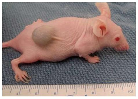
Figure 1 Showing a heterotropic colonic tumor on the flank of the mouse.
(Image used from author’s personal collection).
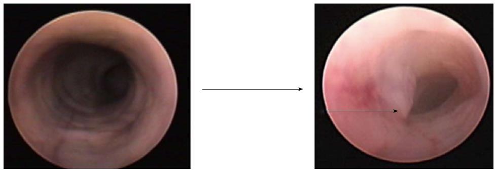
Figure 2 Colonoscopic picture on the left show a normal murine colon with smooth circumferential mucosa.
While the colonoscopic picture on the right shows the orthotopic tumor growth (marked). (Image used from author’s personal collection).
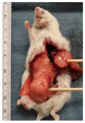
Figure 3 Showing the large orthotopic colon cancer tumor involving the surrounding structures (Lower marker).
Also shown is the metastatic matted group of lymph nodes (Upper marker). (Image used from author’s personal collection).
Figure 4 Showing the orthotopic colon tumor growing from the colonic mucosal surface (Black marker) (hematoxylin-eosin staining, magnification × 100).
There is surrounding normal colonic mucosa (white marker). Adapted from Ref. [20].
- Citation: Mittal VK, Bhullar JS, Jayant K. Animal models of human colorectal cancer: Current status, uses and limitations. World J Gastroenterol 2015; 21(41): 11854-11861
- URL: https://www.wjgnet.com/1007-9327/full/v21/i41/11854.htm
- DOI: https://dx.doi.org/10.3748/wjg.v21.i41.11854


