Copyright
©The Author(s) 2015.
World J Gastroenterol. Sep 7, 2015; 21(33): 9822-9826
Published online Sep 7, 2015. doi: 10.3748/wjg.v21.i33.9822
Published online Sep 7, 2015. doi: 10.3748/wjg.v21.i33.9822
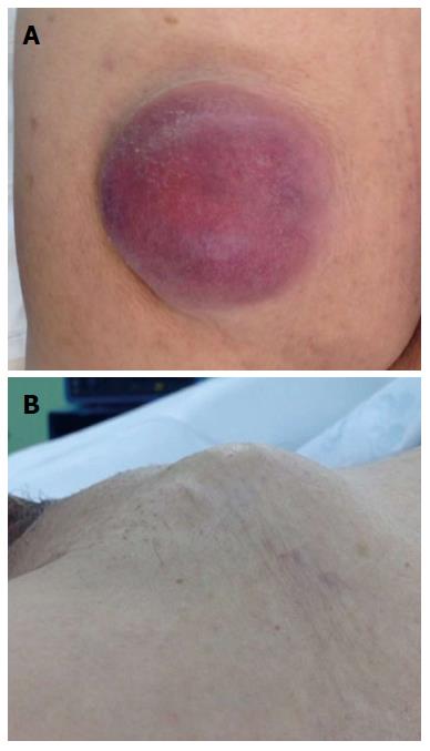
Figure 1 Gross appearance of the left hip mass (A) and the left inguinal mass (B).
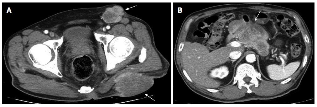
Figure 2 Abdominal computerized tomography scan revealed not only the left hip and inguinal masses (A), but also a heterogeneous enhancing mass on the body of the pancreas (B).
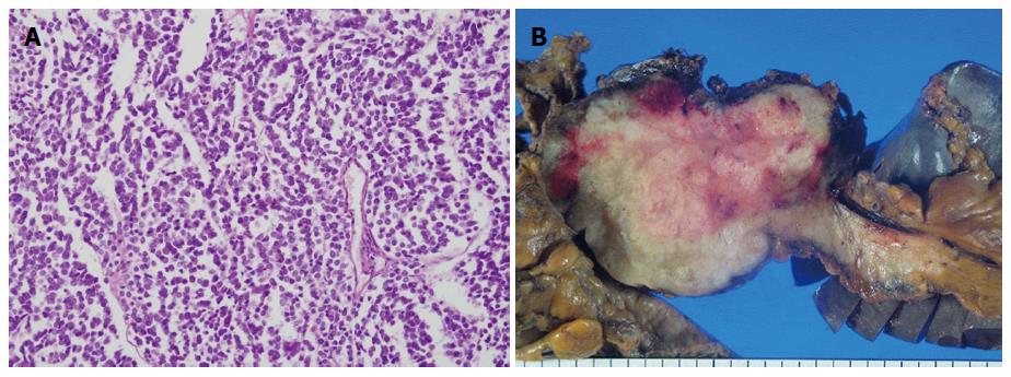
Figure 3 Hematoxylin and eosin stain demonstrated the small to medium-sized cells with scanty cytoplasm and hyperchromatic nucleoli (A, magnification × 200), and the gross appearance of the pancreatic mass (B).
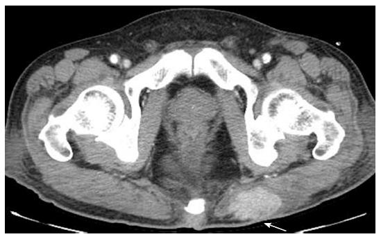
Figure 4 Abdominal computerized tomography scan reveals a 4.
7 cm-sized enhancing mass at the site of the previous mass excision.
- Citation: Shin WY, Lee KY, Ahn SI, Park SY, Park KM. Cutaneous metastasis as an initial presentation of a non-functioning pancreatic neuroendocrine tumor. World J Gastroenterol 2015; 21(33): 9822-9826
- URL: https://www.wjgnet.com/1007-9327/full/v21/i33/9822.htm
- DOI: https://dx.doi.org/10.3748/wjg.v21.i33.9822


