Copyright
©The Author(s) 2015.
World J Gastroenterol. Sep 7, 2015; 21(33): 9817-9821
Published online Sep 7, 2015. doi: 10.3748/wjg.v21.i33.9817
Published online Sep 7, 2015. doi: 10.3748/wjg.v21.i33.9817
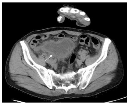
Figure 1 Enhanced computed tomography.
The abdominal scan showed a periappendiceal abscess (arrow).
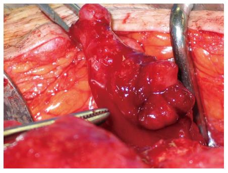
Figure 2 Surgical findings.
The appendix of the patient was diffusely thickened and inflamed, with perforation.
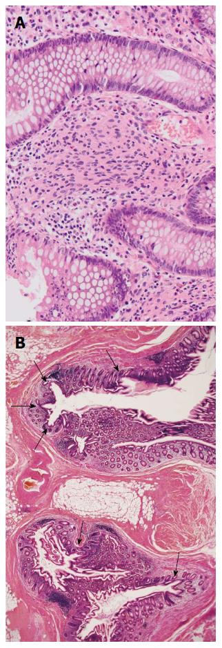
Figure 3 Pathological findings (Hematoxylin-eosin staining).
A: Spindle shaped tumor cells proliferated diffusely in the interstitium between the crypts, without forming a distinct mass (magnification × 10); B: Multiple diverticulums were present in the wall of the appendix. The majority of diverticulums were small in size, causing subtle epithelial herniation (magnification × 1).
- Citation: Ozaki A, Tsukada M, Watanabe K, Tsubokura M, Kato S, Tanimoto T, Kami M, Ohira H, Kanazawa Y. Perforated appendiceal diverticulitis associated with appendiceal neurofibroma in neurofibromatosis type 1. World J Gastroenterol 2015; 21(33): 9817-9821
- URL: https://www.wjgnet.com/1007-9327/full/v21/i33/9817.htm
- DOI: https://dx.doi.org/10.3748/wjg.v21.i33.9817


