Copyright
©The Author(s) 2015.
World J Gastroenterol. Aug 21, 2015; 21(31): 9448-9452
Published online Aug 21, 2015. doi: 10.3748/wjg.v21.i31.9448
Published online Aug 21, 2015. doi: 10.3748/wjg.v21.i31.9448
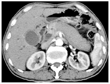
Figure 1 Computed tomography of the enlarged sausage-like pancreas and heterogeneous enhancement of the pancreas in March 2012 prior to steroid treatment.
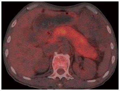
Figure 2 Positron emission tomography/computed tomography showing increased uneven metabolism of the entire pancreas in March 2012.
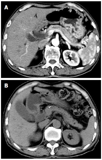
Figure 3 Computed tomography in June and November 2012 revealing the normalized pancreas after steroid treatment.
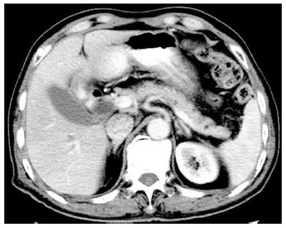
Figure 4 Computed tomography in August 2014 also showing the normal pancreas.
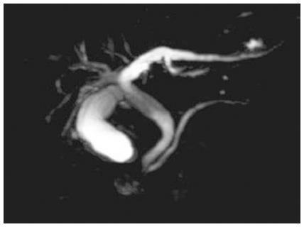
Figure 5 Magnetic resonance cholangiopancreatography showing stenosis of the distal common bile duct and proximal main pancreatic duct, and dilation of the proximal common bile duct and extra- and intra-hepatic bile ducts.
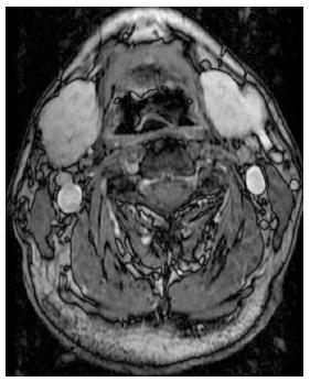
Figure 6 Magnetic resonance imaging of the salivary glands showing symmetrical bilateral enlargement of the submandibular gland (4.
0 × 2.6 × 5.2 cm).
- Citation: Fan RY, Sheng JQ. Immunoglobulin G4-related autoimmune pancreatitis and sialadenitis: A case report. World J Gastroenterol 2015; 21(31): 9448-9452
- URL: https://www.wjgnet.com/1007-9327/full/v21/i31/9448.htm
- DOI: https://dx.doi.org/10.3748/wjg.v21.i31.9448


