Copyright
©The Author(s) 2015.
World J Gastroenterol. Aug 14, 2015; 21(30): 9223-9227
Published online Aug 14, 2015. doi: 10.3748/wjg.v21.i30.9223
Published online Aug 14, 2015. doi: 10.3748/wjg.v21.i30.9223
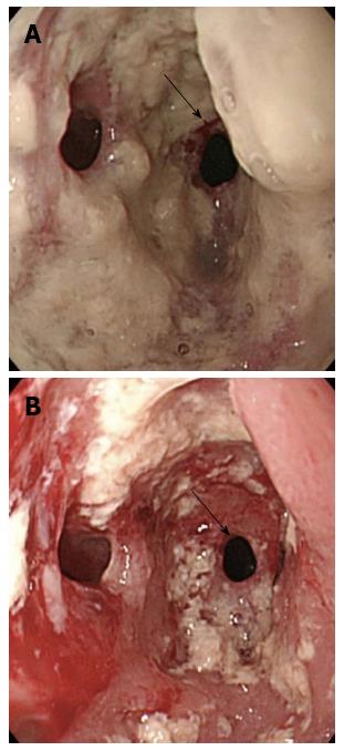
Figure 1 Endoscopy showing an anastomotic stricture with multiple diverticula (arrow) and numerous white plaques (arrow).
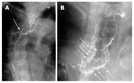
Figure 2 Radiological examination revealing an anastomotic stricture (arrow) (A) and numerous fine, gastrografin-filled projections (arrow) (B).
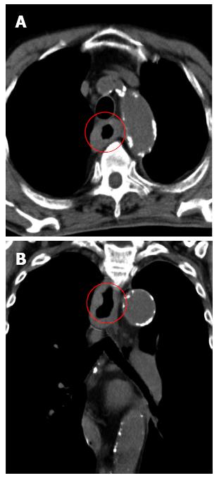
Figure 3 Computed tomography imaging showing the thickness of the wall of the residual esophagus without any sign of metastatic lesions.
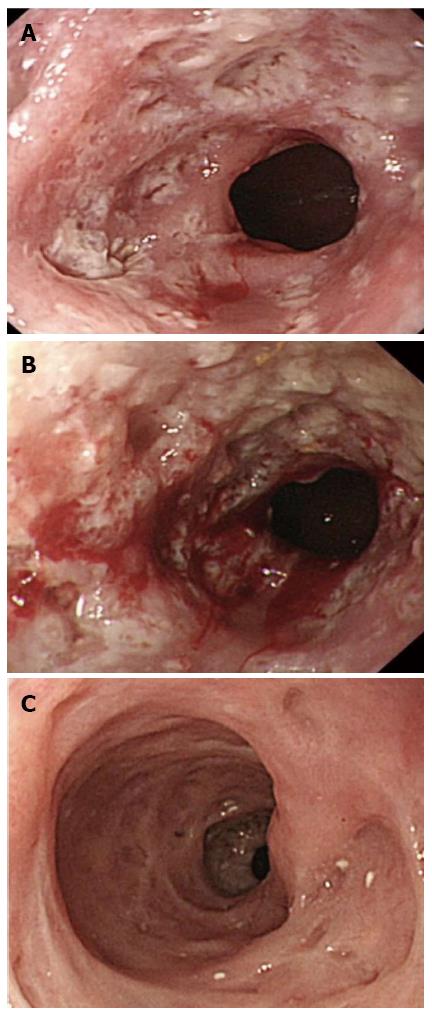
Figure 4 Endoscopic examination.
A: Endoscopic examination revealed that the dilated anastomosis remained and the mucosal inflammation was partially improved; B: Three weeks after discharge, endoscopic examination revealed that the dilated anastomosis remained, while the mucosal inflammation had worsened; C: Two weeks after readmission, endoscopic examination revealed improvements in the mucosal inflammation. Only tiny orifices, corresponding to pseudodiverticulosis, remained.
- Citation: Takeshita N, Kanda N, Fukunaga T, Kimura M, Sugamoto Y, Tasaki K, Uesato M, Sazuka T, Maruyama T, Aida N, Tamachi T, Hosokawa T, Asai Y, Matsubara H. Esophageal intramural pseudodiverticulosis of the residual esophagus after esophagectomy for esophageal cancer. World J Gastroenterol 2015; 21(30): 9223-9227
- URL: https://www.wjgnet.com/1007-9327/full/v21/i30/9223.htm
- DOI: https://dx.doi.org/10.3748/wjg.v21.i30.9223


