Copyright
©The Author(s) 2015.
World J Gastroenterol. Aug 7, 2015; 21(29): 8974-8980
Published online Aug 7, 2015. doi: 10.3748/wjg.v21.i29.8974
Published online Aug 7, 2015. doi: 10.3748/wjg.v21.i29.8974
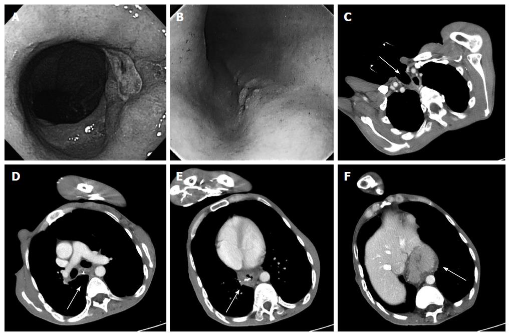
Figure 1 Endoscopy findings (A, B) and computed tomography findings (C-F).
A: An ulcerative lesion was found in a hiatal hernia 30 cm from an incisor; B: 0-IIc type esophageal cancer was detected 25 cm from an incisor; There was severe deformity of the trunk caused by kyphosis and muscular contractures (C-F); C: The arrow points to the permanent tracheal stoma; D: The arrow points to the metal clip near the 0-IIc type esophageal cancer; E: The arrow points to the metal clip near the ulcerative lesion in hiatal hernia; F: The arrow points to the sliding hiatal hernia.
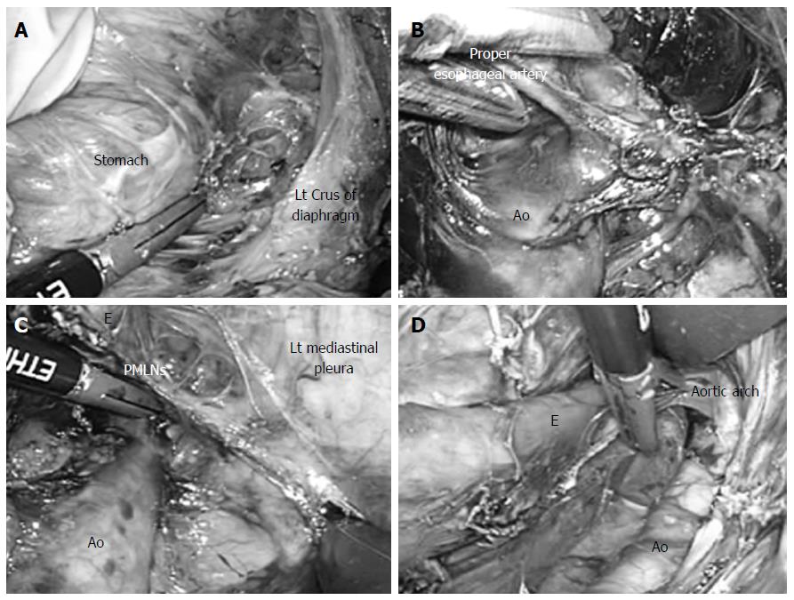
Figure 2 Surgical technique.
A: The esophageal hiatus was divided, and carbon dioxide was introduced into the mediastinum; B: Dissection of the anterior plane of the thoracic aorta was extended to the cranial side, and the root of the proper esophageal artery was confirmed under a magnified videoscopic view; C: While lifting the posterior mediastinal lymph nodes like a membrane, they were cut along the border of the left mediastinal pleura; D: This incision was extended to the left pulmonary hilum and aortic arch. Ao: Thoracic aorta; E: Esophagus; Lt: Left; PMLNs: Posterior mediastinal lymph nodes.
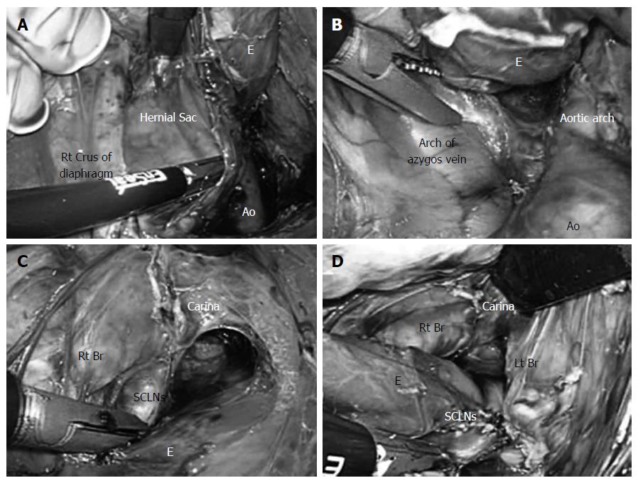
Figure 3 Thoracic esophagus was completely detached from the surrounding tissue.
A: A hernial sac was identified on the cranial side of the right crus of the diaphragm; B: Dissection of the posterior and right sides of the esophagus was performed to the level of the arch of the azygos vein; C: While lifting the right mediastinal pleura like a membrane, an incision was made and extended to the right pulmonary hilum, and the lymph nodes were resected from the right main bronchus and carina; D: Intraoperative view after dissection of the subcarinal lymph nodes. Ao: Thoracic aorta; E: Esophagus; Lt: Left; Rt: Right; Br: Bronchus; SCLNs: Subcarinal lymph nodes.
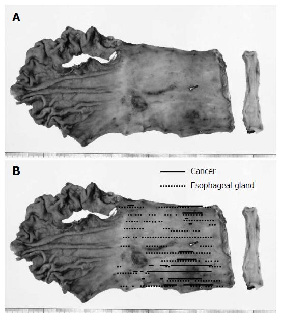
Figure 4 Resected specimen.
A: A resected specimen revealed a 0-IIc type tumor (72 mm × 56 mm in size) in the middle and lower thoracic esophagus in long-segment Barrett's esophagus. A metal clip was identified near the tumor; B: Mapping according to the histopathological examination. Although an Ul-IVs type ulcer (17 mm × 17 mm in size) was identified on the distal side of the tumor, cancer was not found in this lesion.
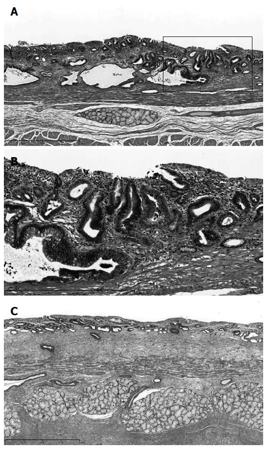
Figure 5 Histopathological examination.
A: A histopathological examination revealed superficial adenocarcinoma (pT1a-SMM). The cancer lesion was surrounded by mucosa, including a columnar epithelium that continued to the stomach; B: Part of the small frame in (A) was magnified and shown; C: Duplication of the muscularis mucosae was identified in long-segment Barrett's esophagus. Hematoxylin-eosin staining; magnification, × 100 (A), × 150 (B) or × 50 (C).
- Citation: Shiozaki A, Fujiwara H, Konishi H, Kinoshita O, Kosuga T, Morimura R, Murayama Y, Komatsu S, Kuriu Y, Ikoma H, Nakanishi M, Ichikawa D, Okamoto K, Sakakura C, Otsuji E. Laparoscopic transhiatal approach for resection of an adenocarcinoma in long-segment Barrett’s esophagus. World J Gastroenterol 2015; 21(29): 8974-8980
- URL: https://www.wjgnet.com/1007-9327/full/v21/i29/8974.htm
- DOI: https://dx.doi.org/10.3748/wjg.v21.i29.8974


