Copyright
©The Author(s) 2015.
World J Gastroenterol. Aug 7, 2015; 21(29): 8804-8810
Published online Aug 7, 2015. doi: 10.3748/wjg.v21.i29.8804
Published online Aug 7, 2015. doi: 10.3748/wjg.v21.i29.8804
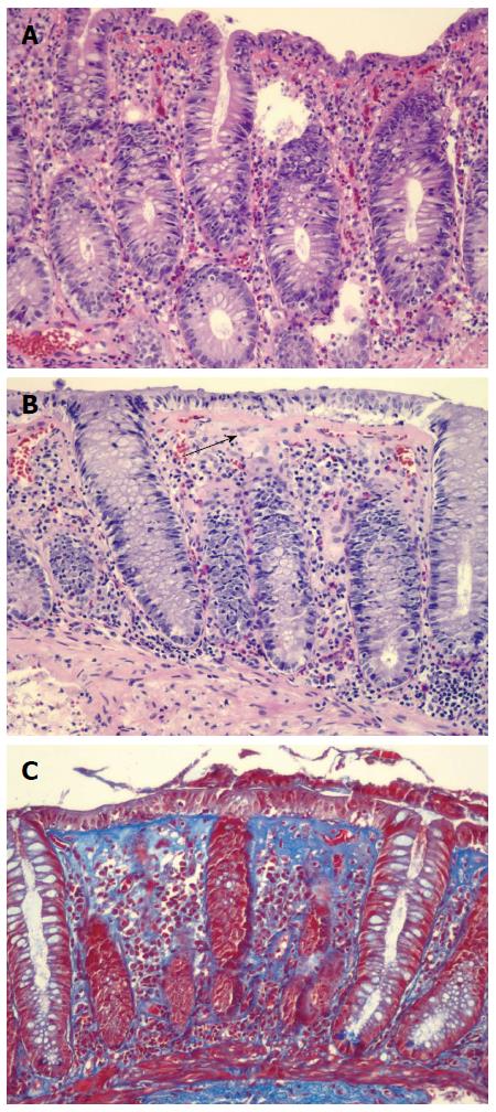
Figure 1 Colonic biopsy.
A: Lymphocytic colitis, hematoxylin and eosin stain. The key histological feature is increased number of intraepithelial lymphocytes with little or no crypt architectural distortion. The presence of > 20 intraepithelial lymphocytes per 100 surface epithelial cells is diagnostic of lymphocytic colitis (LC)[2]. Fewer than 5 intraepithelial lymphocytes per 100 surface epithelial cells are considered normal[2]. Inflammatory infiltration usually consists of lymphocytes and plasma cells but eosinophils and neutrophils may also be present; B: Collagenous colitis, hematoxylin and eosin stain. The black arrow points to the thickened subepithelial collagen band. Band thickness is 10-20 μm in collagenous colitis (CC)[2]. Collagen band thickness < 3 μm is considered normal[2]; C: Collagenous colitis, trichrome stain. Collagen stains blue on trichrome stain. Trichrome stain can be useful when it is difficult to determine the thickness of collagen band on hematoxylin and eosin stain. This picture demonstrates a thickened subepithelial collagen band.
- Citation: Park T, Cave D, Marshall C. Microscopic colitis: A review of etiology, treatment and refractory disease. World J Gastroenterol 2015; 21(29): 8804-8810
- URL: https://www.wjgnet.com/1007-9327/full/v21/i29/8804.htm
- DOI: https://dx.doi.org/10.3748/wjg.v21.i29.8804


