Copyright
©The Author(s) 2015.
World J Gastroenterol. Jul 14, 2015; 21(26): 8118-8124
Published online Jul 14, 2015. doi: 10.3748/wjg.v21.i26.8118
Published online Jul 14, 2015. doi: 10.3748/wjg.v21.i26.8118
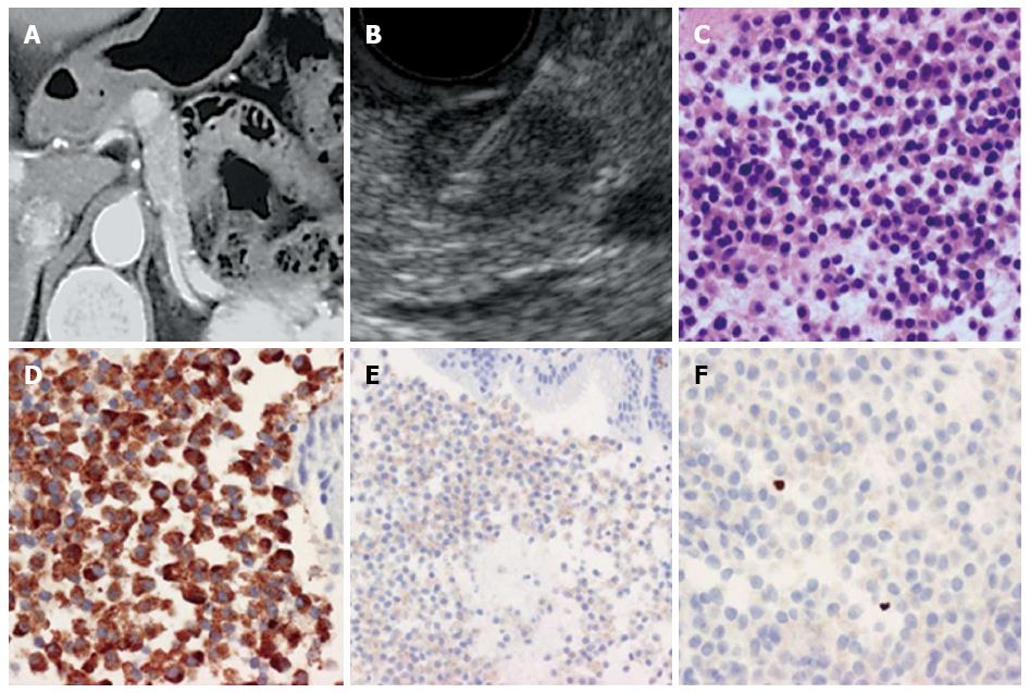
Figure 1 Method of endoscopic ultrasonography-guided fine needle aspiration.
A: Abdominal contrast computed tomography. An enhancing tumor was recognized in the body of the pancreas; B: EUS-FNA. The tumor was recognized as a low echoic lesion with distinct boundaries; a 22G needle was inserted into the tumor; C: A specimen obtained by EUS-FNA (HE staining). Tumor cells with oval nuclei and acidophilic granular cytoplasm proliferated diffusely; D: A specimen obtained by EUS-FNA (Chromogranin A staining). Tumor cells were Chromogranin A-positive; E: A specimen taken by EUS-FNA (CD56 staining). Tumor cells were CD56-positive; F: A specimen taken by EUS-FNA (Ki-67 antibody staining). The Ki-67 index was 0.4% with tumor grade G1. EUS-FNA: Endoscopic ultrasonography-guided fine needle aspiration.
- Citation: Sugimoto M, Takagi T, Hikichi T, Suzuki R, Watanabe K, Nakamura J, Kikuchi H, Konno N, Waragai Y, Asama H, Takasumi M, Watanabe H, Obara K, Ohira H. Efficacy of endoscopic ultrasonography-guided fine needle aspiration for pancreatic neuroendocrine tumor grading. World J Gastroenterol 2015; 21(26): 8118-8124
- URL: https://www.wjgnet.com/1007-9327/full/v21/i26/8118.htm
- DOI: https://dx.doi.org/10.3748/wjg.v21.i26.8118


