Copyright
©The Author(s) 2015.
World J Gastroenterol. Jul 7, 2015; 21(25): 7764-7776
Published online Jul 7, 2015. doi: 10.3748/wjg.v21.i25.7764
Published online Jul 7, 2015. doi: 10.3748/wjg.v21.i25.7764
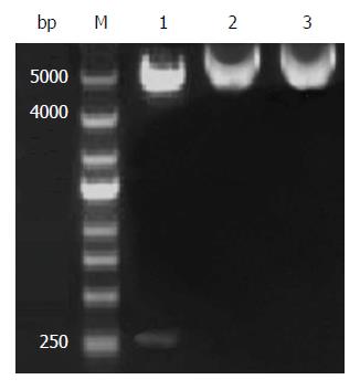
Figure 1 Restriction analysis of pEGFP-N1- high mobility group box 1 protein A-box.
Lane M: marker; lane 1: pEGFP-N1- high mobility group box 1 protein (HMGB1) A-box digested by XhoI + BamHI; lane 2: pEGFP-N1-HMGB1 A-box digested by XhoI; and lane 3: pEGFP-N1-HMGB1 A-box digested by BamHI.
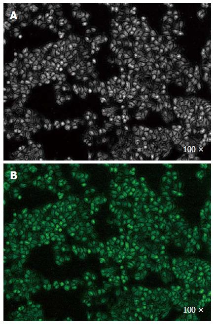
Figure 2 SW480 cells at 48 h after transfection (magnification × 100).
A: Bright-field images of cells (after 48 h); B: Green fluorescence of transfected cells (after 48 h).
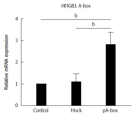
Figure 3 Expression of HMGB1 protein pA-box mRNA in transfected SW480 cells.
After SW480 cells were transfected for 48 h, HMGB1 pA-box mRNA levels were determined by real-time PCR. The expression of HMGB1 A-box mRNA in the pA-box-transfection SW480 cells was significantly upregulated compared with the control and mock-transfection groups. The data are expressed as the mean ± SD and are derived from three independent experiments, which were each performed in duplicate. The means with asterisks above them are significantly different (bP < 0.01 vs control).
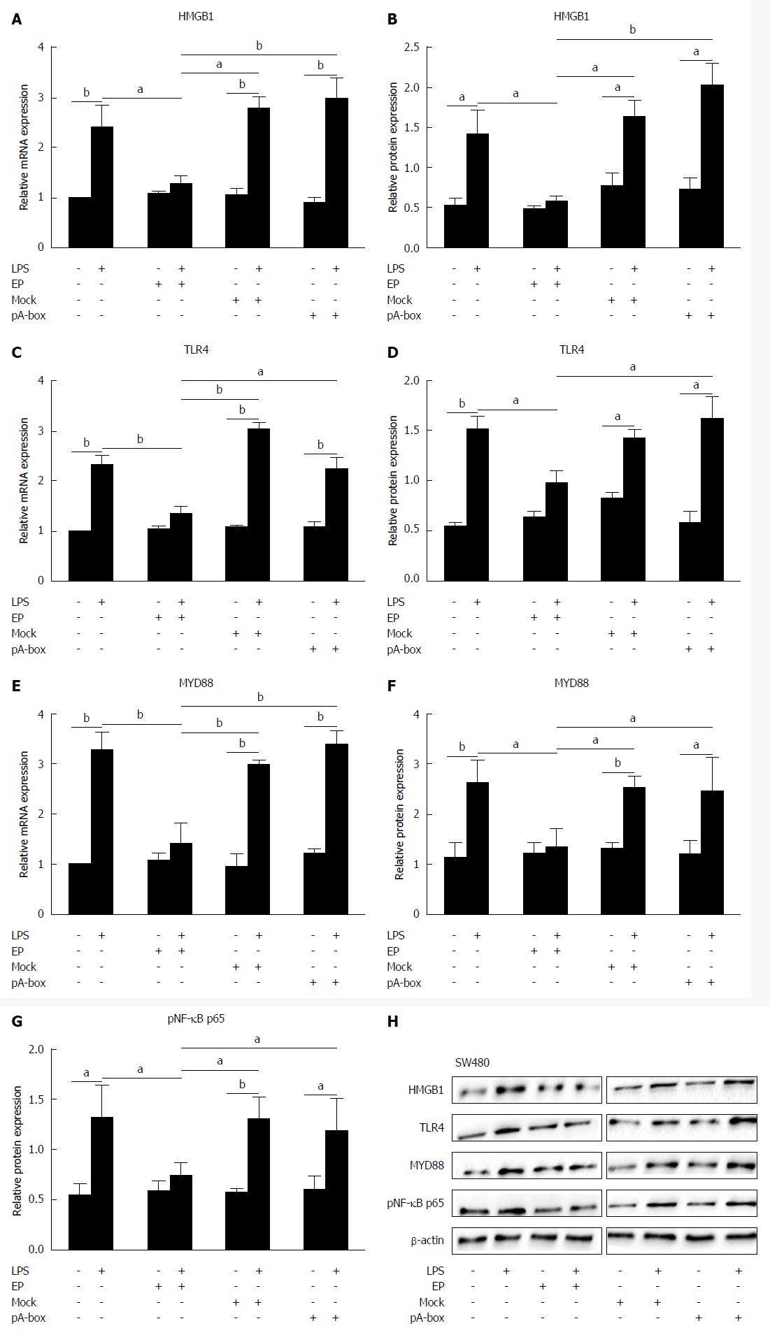
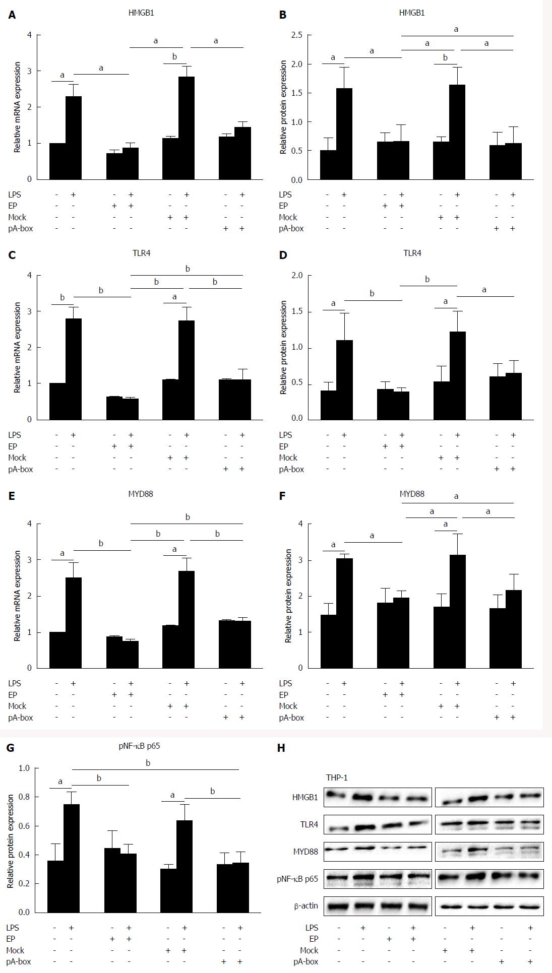
Figure 4 Expression of the HMGB1 protein and toll-like receptor 4 signaling pathways in lipopolysaccharide-stimulated SW480 cells.
After pretreatment with EP (5 mmol/L, 1 h), the SW480 cells were treated with LPS (1 μg/mL, 24 h). The HMGB1 protein, TLR4, MYD88, and pNF-κB p65 mRNA and protein levels were determined by real-time PCR (A, C, E), densitometric quantification and representative western blotting with β-actin as the loading control (B, D, F, G, H). LPS increased the HMGB1, TLR4, MYD88, and pNF-κB p65 mRNA and protein levels in the LPS-stimulated SW480 cells compared with the LPS-untreated group, but EP downregulated these mRNA and protein levels in the LPS-stimulated SW480 cells. By contrast, HMGB1 A-box failed to downregulate the HMGB1, TLR4, MYD88, and pNF-κB p65 mRNA and protein levels compared with the mock-transfection group. The data are expressed as the mean ± SD and are derived from three independent experiments, which were each performed in duplicate. The means with letters above them are significantly different (aP < 0.05, bP < 0.01 vs control).
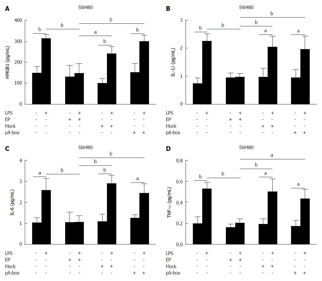
Figure 5 Levels of the inflammatory mediators HMGB1 protein, IL-1β, IL-6 and TNF-α in the SW480 cell supernatant.
After pretreatment with EP (5 mmol/L, 1 h), the SW480 cells were treated with LPS (1 μg/mL, 24 h). The levels of the inflammatory mediators HMGB1, IL-1β, IL-6 and TNF-α in the supernatant of the SW480 cells were detected by ELISA (A-D). Compared with the LPS-untreated group, LPS increased the levels of HMGB1, IL-1β, IL-6 and TNF-α in the LPS-stimulated SW480 cells, but EP downregulated the levels of HMGB1, IL-1β, IL-6 and TNF-α in the SW480 cells activated by LPS. By contrast, HMGB1 A-box failed to decrease the secretion of HMGB1, IL-1β, IL-6 and TNF-α compared with the mock-transfection group. The data are expressed as the mean ± SD and are derived from three independent experiments, which were each performed in duplicate. The means with letters above them are significantly different (aP < 0.05, bP < 0.01 vs control).
Figure 6 Expression of the HMGB1 protein and TLR4 signaling pathways in THP-1 cells co-cultured with SW480 cells.
After pretreatment with EP (5 mmol/L, 1 h), the SW480 cells were treated with 1 μg/mL LPS and co-cultured with THP-1 cells for 24 h. The HMGB1, TLR4, MYD88, pNF-κB p65 mRNA and protein levels were determined by real-time PCR (A, C, E), densitometric quantification and representative western blots, with β-actin as the loading control (B, D, F, G, H). Compared with the LPS-untreated group, LPS increased HMGB1, TLR4, MYD88, pNF-κB p65 mRNA and protein levels in the THP-1 cells, but EP downregulated these levels. Similarly, HMGB1 A-box also downregulated HMGB1, TLR4, MYD88, pNF-κB p65 mRNA and protein levels compared with the mock-transfected group. The data are expressed as the mean ± SD and are derived from three independent experiments, which were each performed in duplicate. The means with letters above them are significantly different (aP < 0.05, bP < 0.01 vs control).
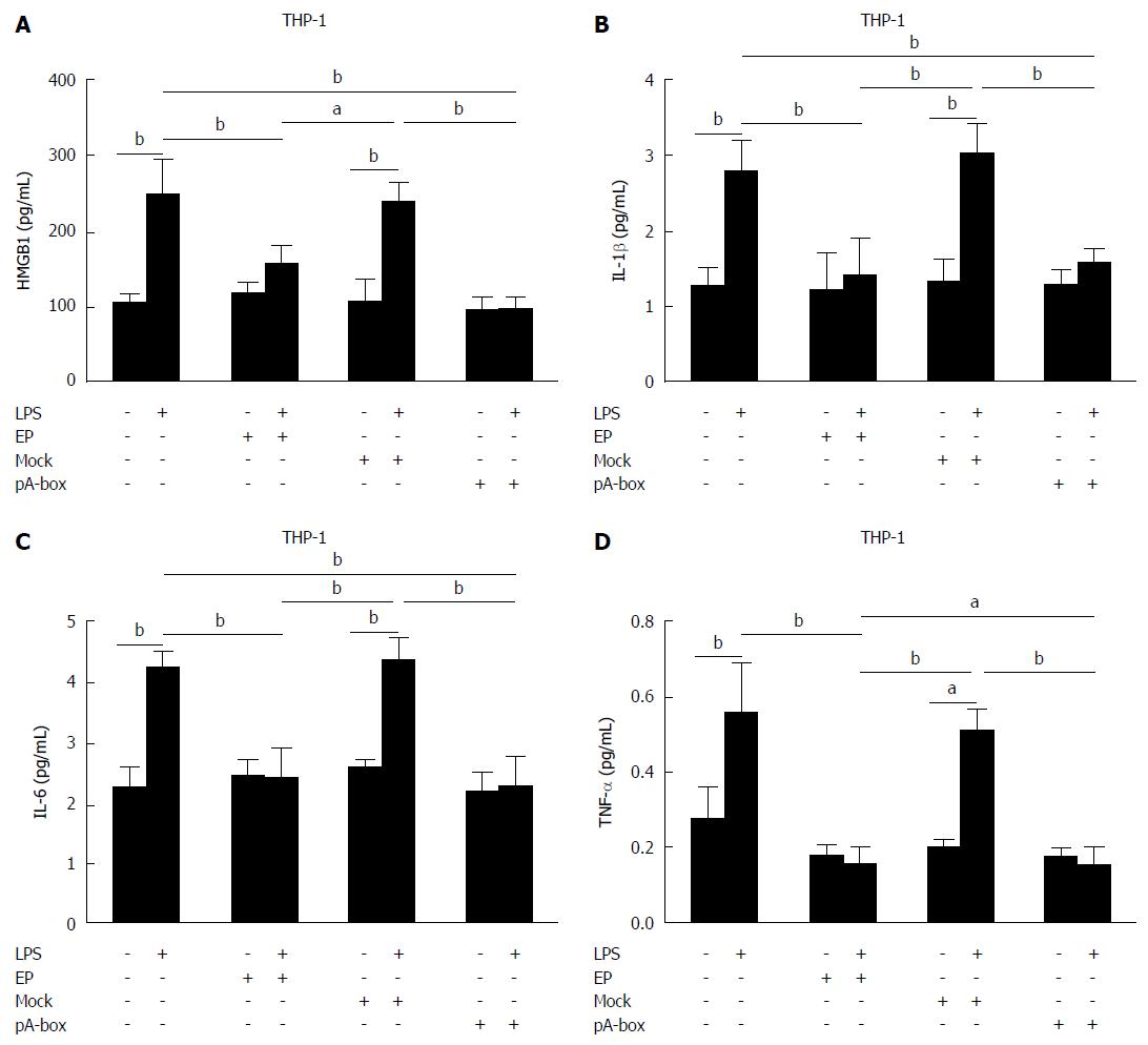
Figure 7 Levels of the inflammatory mediators high mobility group box 1 protein, IL-1β, IL-6 and TNF-α in the supernatant of the THP-1 cells co-cultured with SW480 cells.
After pretreatment with EP (5 mmol/L, 1 h), the SW480 cells were treated with 1 μg/mL LPS and co-cultured with THP-1 cells for 24 h. The levels of the inflammatory mediators HMGB1, IL-1β, IL-6 and TNF-α in the supernatant of the THP-1 cells co-cultured with SW480 cells were detected by ELISA (A-D). Compared with the LPS-untreated group, LPS increased the levels of HMGB1, IL-1β, IL-6 and TNF-α in the THP-1 cells, but EP downregulated these levels. Similarly, HMGB1 A-box also decreased the secretion of HMGB1, IL-1β, IL-6 and TNF-α in the THP-1 cells compared with the mock-transfection group. The data are expressed as the mean ± SD and are derived from three independent experiments, which were each performed in duplicate. The means with letters above them are significantly different (aP < 0.05, bP < 0.01 vs control).
-
Citation: Wang FC, Pei JX, Zhu J, Zhou NJ, Liu DS, Xiong HF, Liu XQ, Lin DJ, Xie Y. Overexpression of HMGB1 A-box reduced lipopolysaccharide-induced intestinal inflammation
via HMGB1/TLR4 signalingin vitro . World J Gastroenterol 2015; 21(25): 7764-7776 - URL: https://www.wjgnet.com/1007-9327/full/v21/i25/7764.htm
- DOI: https://dx.doi.org/10.3748/wjg.v21.i25.7764


