Copyright
©The Author(s) 2015.
World J Gastroenterol. Jul 7, 2015; 21(25): 7683-7708
Published online Jul 7, 2015. doi: 10.3748/wjg.v21.i25.7683
Published online Jul 7, 2015. doi: 10.3748/wjg.v21.i25.7683
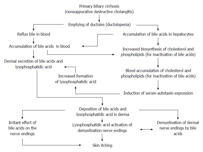
Figure 1 Scheme of mechanism of development of pruritus in primary biliary cirrhosis.
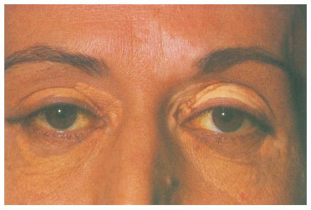
Figure 2 Xanthelasmas on both eye lids in a patient with primary biliary cirrhosis.
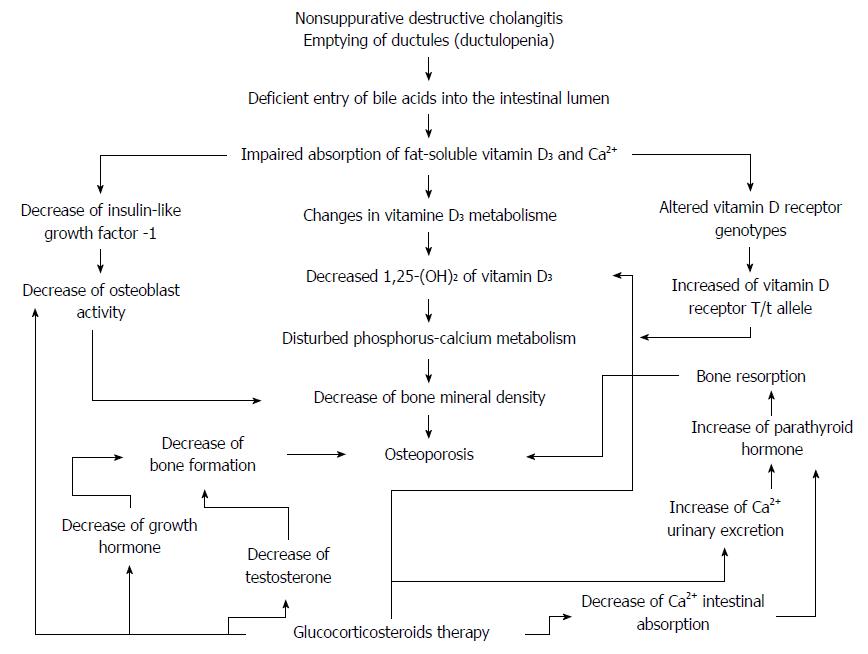
Figure 3 The scheme of the development of osteoporosis in primary biliary cirrhosis.
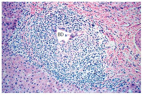
Figure 4 Stage I primary biliary cirrhosis.
Inflammatory infiltrate consisting of mononuclear cells around the bile duct (BD). Hematoxylin eosin staining, magnification × 125.
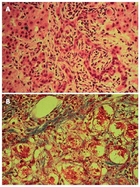
Figure 5 Stages II-III primary biliary cirrhosis.
A: Wide septa formed by proliferating ductuli. Lymphocytic infiltration. Hematoxylin eosin staining, magnification × 200. B: Hyaline inclusions. Swelling and hepatocellular injury due to the damaging effect of bile acids. van Gieson's, magnification × 200.
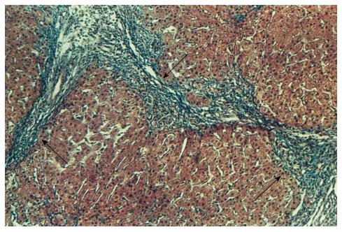
Figure 6 Stages IV primary biliary cirrhosis.
Cirrhotic transformation. Silver impregnation method for Gomory, magnification × 200.
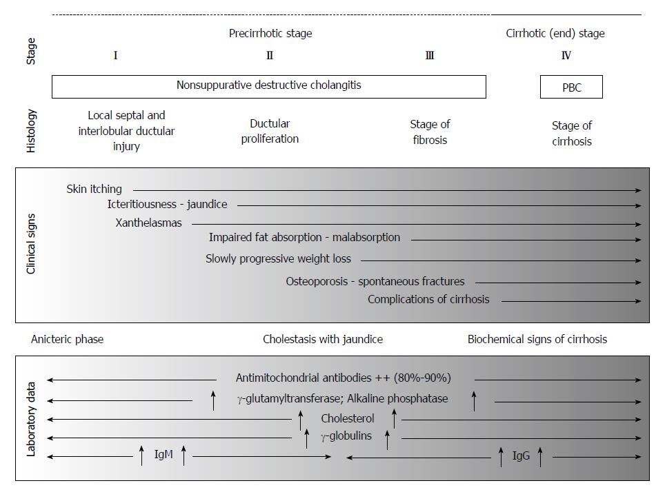
Figure 7 Schematically summarizes the clinical, biochemical, immunological, and morphological signs of primary biliary cirrhosis.
- Citation: Reshetnyak VI. Primary biliary cirrhosis: Clinical and laboratory criteria for its diagnosis. World J Gastroenterol 2015; 21(25): 7683-7708
- URL: https://www.wjgnet.com/1007-9327/full/v21/i25/7683.htm
- DOI: https://dx.doi.org/10.3748/wjg.v21.i25.7683


