Copyright
©The Author(s) 2015.
World J Gastroenterol. Jul 7, 2015; 21(25): 7659-7671
Published online Jul 7, 2015. doi: 10.3748/wjg.v21.i25.7659
Published online Jul 7, 2015. doi: 10.3748/wjg.v21.i25.7659
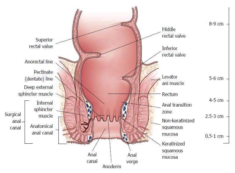
Figure 1 Rectal anatomy and landmarks of importance in the treatment of rectal cancer (Figure reproduced with permission from Apgar et al[3]).
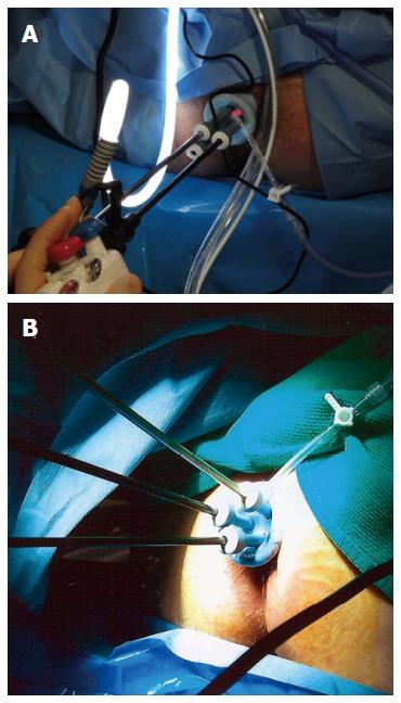
Figure 2 Operative setup for transanal minimally invasive surgery (Figure reproduced with permission from Atallah et al[67]).
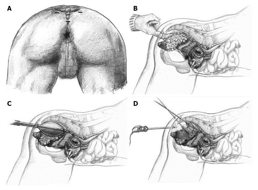
Figure 3 Technique of endoscopic posterior mesorectal resection.
A: Trocar positions; B: Access to the retrorectal space using the index finger; C: Establishment of a sufficient large operating space using a dissecting balloon trocar; D: Dissection of the mesorectum from the posterior wall of the rectum. Figure reproduced with permission from Zerz et al[70].
- Citation: Gaertner WB, Kwaan MR, Madoff RD, Melton GB. Rectal cancer: An evidence-based update for primary care providers. World J Gastroenterol 2015; 21(25): 7659-7671
- URL: https://www.wjgnet.com/1007-9327/full/v21/i25/7659.htm
- DOI: https://dx.doi.org/10.3748/wjg.v21.i25.7659


