Copyright
©The Author(s) 2015.
World J Gastroenterol. Jun 21, 2015; 21(23): 7181-7190
Published online Jun 21, 2015. doi: 10.3748/wjg.v21.i23.7181
Published online Jun 21, 2015. doi: 10.3748/wjg.v21.i23.7181
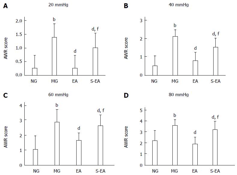
Figure 1 Comparison of the abdominal withdrawal reflex scores of the rats in each group.
Under colorectal distension stimulation at the same strength; bP < 0.01 vs NG; dP < 0.01 vs MG; fP < 0.01 vs EA. NG: Normal group; MG: Model group; EA: Electroacupuncture group; S-EA: Sham EA group; AWR: Abdominal withdrawal reflex.
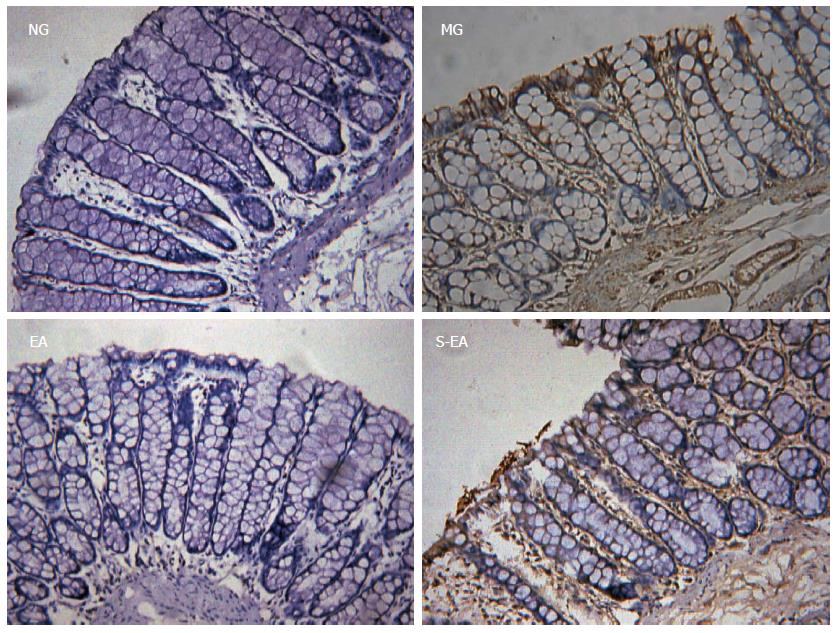
Figure 2 Expression of corticotropin-releasing hormone in the colon tissue of the rats in each group (magnification × 200).
NG: Normal group; MG: Model group; EA: Electroacupuncture group; S-EA: Sham EA group.
Figure 3 Corticotropin-releasing hormone content in the spinal cord and hypothalamus of the rats in each group.
aP < 0.05 vs NG; bP < 0.01 vs MG; cP < 0.05 vs EA. NG: Normal group; MG: Model group; EA: Electroacupuncture group; S-EA: Sham EA group.
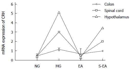
Figure 4 mRNA expression of corticotropin-releasing hormone in the colon, spinal cord, and hypothalamus of the rats in each group.
NG: Normal group; MG: Model group; EA: Electroacupuncture group; S-EA: Sham EA group.
- Citation: Liu HR, Fang XY, Wu HG, Wu LY, Li J, Weng ZJ, Guo XX, Li YG. Effects of electroacupuncture on corticotropin-releasing hormone in rats with chronic visceral hypersensitivity. World J Gastroenterol 2015; 21(23): 7181-7190
- URL: https://www.wjgnet.com/1007-9327/full/v21/i23/7181.htm
- DOI: https://dx.doi.org/10.3748/wjg.v21.i23.7181


