Copyright
©The Author(s) 2015.
World J Gastroenterol. Jun 14, 2015; 21(22): 7047-7051
Published online Jun 14, 2015. doi: 10.3748/wjg.v21.i22.7047
Published online Jun 14, 2015. doi: 10.3748/wjg.v21.i22.7047
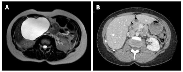
Figure 1 Magnetic resonance imaging and post-operative computed tomography scan of patient 1.
Magnetic resonance imaging shows a large serous cystadenoma in the pancreatic head measured at 13 cm (A), with post-operative computed tomography scan showing complete disappearance after laparoscopic fenestration (B).
Figure 2 Computed tomography scan and post-operative magnetic resonance imaging of patient 2.
Computed tomography scan showed a serous cystadenoma in the pancreatic head measured at 4 cm at diagnosis (A) and 12 cm (B) before surgery; post-operative magnetic resonance imaging showing complete cyst disappearance (C).
Figure 3 Magnetic resonance imaging and post-operative computed tomography scan of patient 3.
Magnetic resonance imaging showing multiple serous cystadenoma, with a large one in the pancreatic head measuring at 10 cm at diagnosis (A) and 14 cm before surgery (B); post-operative computed tomography scan showed complete disappearance (C).
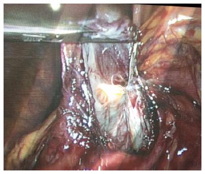
Figure 4 Intraoperative view, showing large fenestration of a large serous cystadenoma in the pancreatic head.
- Citation: Dokmak S, Aussilhou B, Rasoaherinomenjanahary F, Sauvanet A, Vullierme MP, Rebours V, Lévy P. Laparoscopic fenestration of pancreatic serous cystadenoma: Minimally invasive approach for symptomatic benign disease. World J Gastroenterol 2015; 21(22): 7047-7051
- URL: https://www.wjgnet.com/1007-9327/full/v21/i22/7047.htm
- DOI: https://dx.doi.org/10.3748/wjg.v21.i22.7047


