Copyright
©The Author(s) 2015.
World J Gastroenterol. Jun 14, 2015; 21(22): 6944-6951
Published online Jun 14, 2015. doi: 10.3748/wjg.v21.i22.6944
Published online Jun 14, 2015. doi: 10.3748/wjg.v21.i22.6944
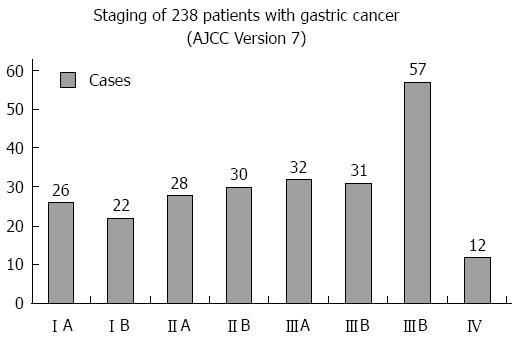
Figure 1 Staging of 238 patients with gastric cancer (AJCC Version 7).
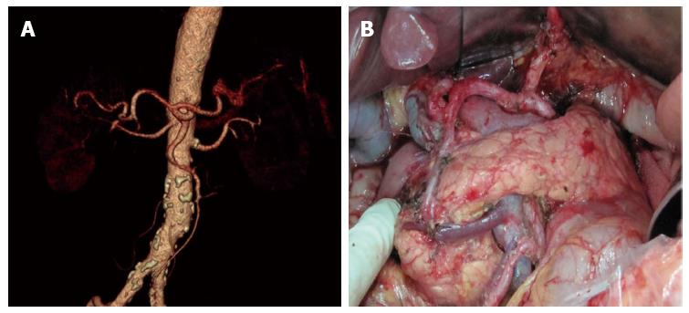
Figure 2 Normal celiac artery branches.
A: Preoperative multi-slice computed tomography angiography: Normal celiac trunk gives off LGA, splenic artery and common hepatic artery; B: Intraoperative finding was consistent with preoperative findings.
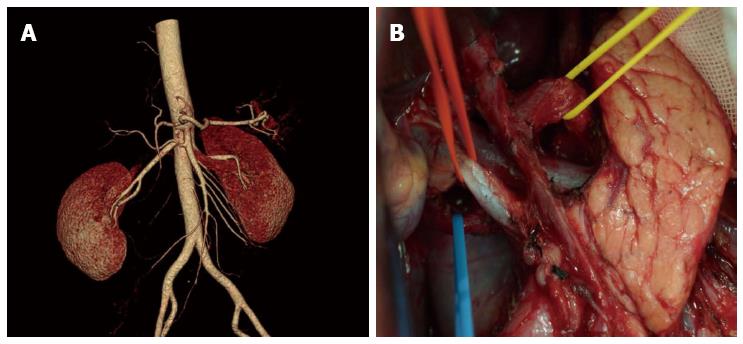
Figure 3 Replaced right hepatic artery.
A: Preoperative multi-slice computed tomography angiography: Replaced right hepatic artery deriving from superior mesenteric artery; B: Intraoperative finding: Replaced right hepatic artery deriving from superior mesenteric artery in the same patient.
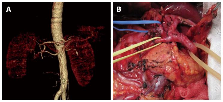
Figure 4 Left gastric artery absence.
A: Preoperative multi-slice computed tomography angiography found left gastric artery absence; B: Intraoperative dissection did not find left gastric artery existence in the same patient.
- Citation: Huang Y, Mu GC, Qin XG, Chen ZB, Lin JL, Zeng YJ. Study of celiac artery variations and related surgical techniques in gastric cancer. World J Gastroenterol 2015; 21(22): 6944-6951
- URL: https://www.wjgnet.com/1007-9327/full/v21/i22/6944.htm
- DOI: https://dx.doi.org/10.3748/wjg.v21.i22.6944


