Copyright
©The Author(s) 2015.
World J Gastroenterol. Jun 7, 2015; 21(21): 6764-6768
Published online Jun 7, 2015. doi: 10.3748/wjg.v21.i21.6764
Published online Jun 7, 2015. doi: 10.3748/wjg.v21.i21.6764
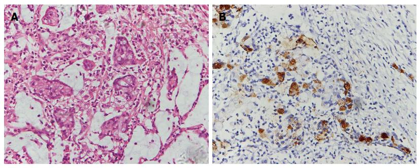
Figure 1 Photomicrographs show hyperchromatic and pleomorphic neoplastic cells of the signet ring type floating in the mucinous lakes among the gastric cancer tissue (hematoxylin and eosin staining, magnification × 40) (A) immunoreactivity of the tumor cells to MUC-5 (magnification × 40) (B).
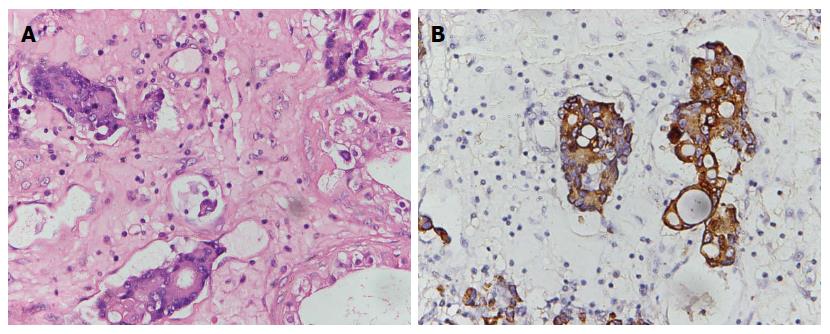
Figure 2 Photomicrographs show the infiltration of the paratesticular tissue by metastatic poorly differentiated adenocarcinoma cells: (hematoxylin and eosin staining, magnification × 40) (A), immunoreactivity of the tumor cells to CAM5.
2 (magnification × 40) (B).
- Citation: Li B, Cai H, Kang ZC, Wu H, Hou JG, Ma LY. Testicular metastasis from gastric carcinoma: A case report. World J Gastroenterol 2015; 21(21): 6764-6768
- URL: https://www.wjgnet.com/1007-9327/full/v21/i21/6764.htm
- DOI: https://dx.doi.org/10.3748/wjg.v21.i21.6764


