Copyright
©The Author(s) 2015.
World J Gastroenterol. Jun 7, 2015; 21(21): 6754-6758
Published online Jun 7, 2015. doi: 10.3748/wjg.v21.i21.6754
Published online Jun 7, 2015. doi: 10.3748/wjg.v21.i21.6754
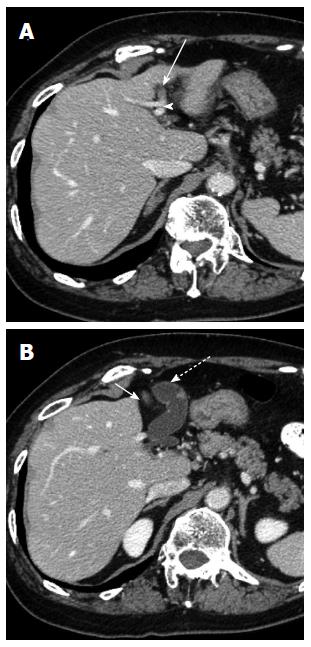
Figure 1 Computed tomography examination reveals a left-sided gallbladder.
A: The round ligament (arrow) is connected to the left portal umbilical portion (arrow head); B: The gallbladder (dotted arrow) is located to the left side of the round ligament (arrow) and the middle hepatic vein, and attached to the left lateral section of the liver.
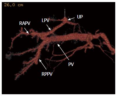
Figure 2 Computed tomography portography reveals the portal venous anomaly.
The dotted line is the round ligament. PV: Portal vein; LPV: Left portal vein; RAPV: Right anterior portal vein; RPPV: Right posterior portal vein; UP: Umbilical portion.
Figure 3 Drip infusion cholangiographic-computed tomography demonstrates that the cystic duct joins the extrahepatic bile duct on the right side and creates a hairpin turn anterior to the extrahepatic bile duct.
One of two bile ducts of segment 2 (arrow) follows a route that is caudal to the umbilical portion of the left portal vein, that is, an infraportal course, and joins the extrahepatic bile duct; the other one (dotted arrow) follows a normal route, that is, a supraportal course. A: Computed tomography (CT) three-dimensional reconstruction, ventral view; B: CT three-dimensional reconstruction, dorsal view.
Figure 4 Intraoperative finding reveals a true left-sided gallbladder.
A: Image before cholecystectomy; B: Anomaly of the cholecystic vein which enters the round ligament; C: Image after cholecystectomy.
- Citation: Ishii H, Noguchi A, Onishi M, Takao K, Maruyama T, Taiyoh H, Araki Y, Shimizu T, Izumi H, Tani N, Yamaguchi M, Yamane T. True left-sided gallbladder with variations of bile duct and cholecystic vein. World J Gastroenterol 2015; 21(21): 6754-6758
- URL: https://www.wjgnet.com/1007-9327/full/v21/i21/6754.htm
- DOI: https://dx.doi.org/10.3748/wjg.v21.i21.6754


