Copyright
©The Author(s) 2015.
World J Gastroenterol. Jun 7, 2015; 21(21): 6706-6712
Published online Jun 7, 2015. doi: 10.3748/wjg.v21.i21.6706
Published online Jun 7, 2015. doi: 10.3748/wjg.v21.i21.6706
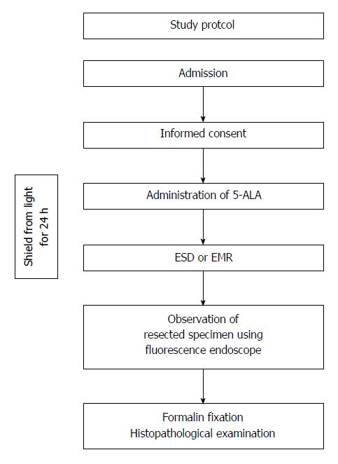
Figure 1 Flow chart of the study.
5-ALA: 5-aminolevulinic acid; ESD: Endoscopic submucosal dissection; EMR: Endoscopic mucosal resection.
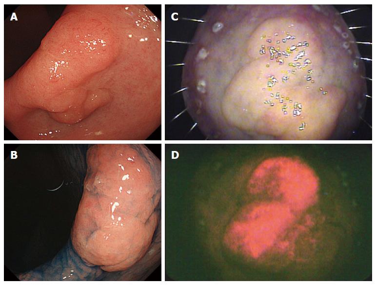
Figure 2 Early gastric cancer with fluorescence.
A: Endoscopic image; B: Endoscopic image obtained after the spraying of indigo carmine dye; C: Resected specimen irradiated by white light; D: Resected specimen showing fluorescence.
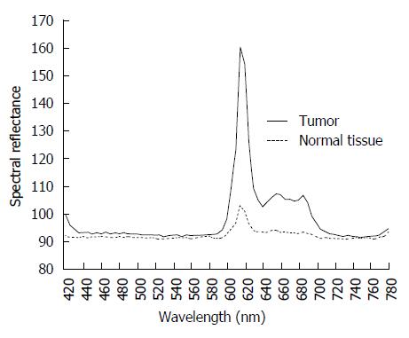
Figure 3 Spectral reflectance in the range of 400-800 nm obtained using a hyperspectral camera.
Two reflectance peaks in the tumor were detected at 635 and 700 nm; this bimodal reflectance is the characteristic reflectance pattern of protoporphyrin IX.
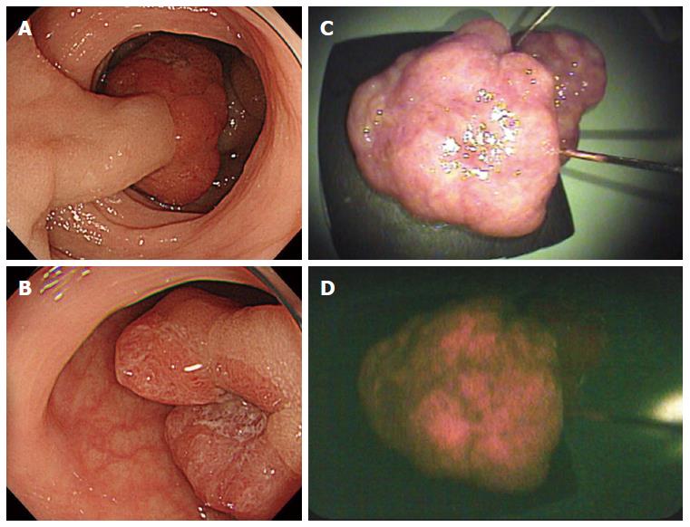
Figure 4 Early colorectal cancer with fluorescence.
A and B: Endoscopic images; C: Resected specimen irradiated by white light; D: Resected specimen showing fluorescence.
- Citation: Nakamura M, Nishikawa J, Hamabe K, Goto A, Nishimura J, Shibata H, Nagao M, Sasaki S, Hashimoto S, Okamoto T, Sakaida I. Preliminary study of photodynamic diagnosis using 5-aminolevulinic acid in gastric and colorectal tumors. World J Gastroenterol 2015; 21(21): 6706-6712
- URL: https://www.wjgnet.com/1007-9327/full/v21/i21/6706.htm
- DOI: https://dx.doi.org/10.3748/wjg.v21.i21.6706


