Copyright
©The Author(s) 2015.
World J Gastroenterol. May 28, 2015; 21(20): 6146-6156
Published online May 28, 2015. doi: 10.3748/wjg.v21.i20.6146
Published online May 28, 2015. doi: 10.3748/wjg.v21.i20.6146
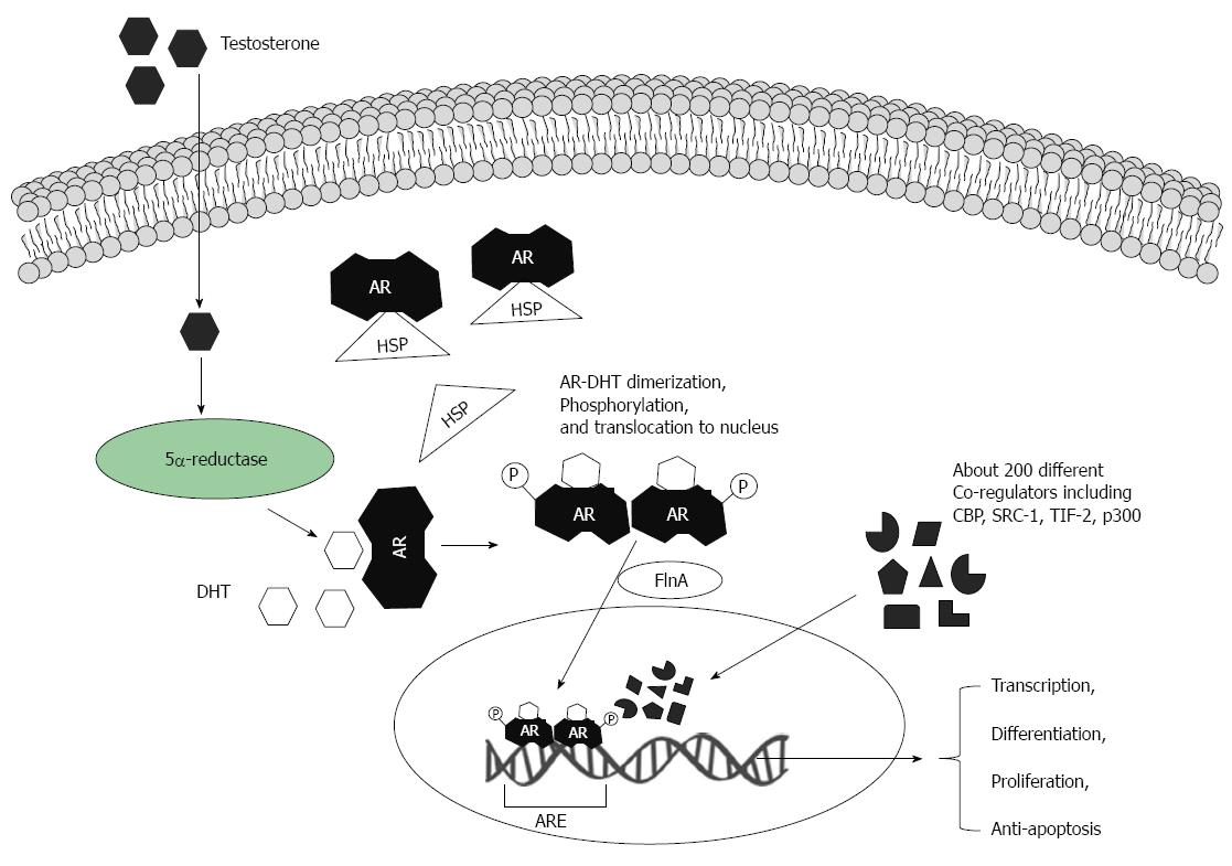
Figure 1 Biological actions of androgens via androgen receptors.
Testosterone molecules translocate via the plasma membrane and are transformed into dihydrotestosterone (DHT) by 5α-reductase. The androgen receptor (AR) is located in cytoplasm and bound to heat shock protein (HSP). DHT binds to the AR and HSP is then released. Ligand-AR complexes can be phosphorylated (and/or are modified by other post-translational mechanisms). Two ligand-AR complexes form homodimers and move into the nucleus. AR nuclear translocation is facilitated by filamin A (FlnA). In the cell nucleus ligand-AR complexes bind to specific DNA elements - androgen-responsive elements (ARE), which are in target gene promoters. These regulate target gene expression at the transcriptional level. A large variety of co-factors and regulators can orchestrate AR-induced gene transcription.
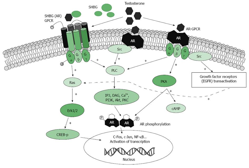
Figure 2 Schematic presentation of testosterone-activated extra-nuclear signaling pathways.
Testosterone binds to undefined membrane-associated androgen receptor(s) (mAR) that might transduce signaling downstream to phospholipase c (PLC). Activation of PLC produces several second messengers including Ins(1,4,5)P3 (IP3) and DAG. Ca2+ influx then leads to an increase in intracellular Ca2+. Alternatively, in the ERK pathway, Testosterone binds to the membrane-associated receptor, which associates with and activates Src kinase. In a third proposed mechanism SHBG-AR GPCR activates Ras, which in turn activates the cascade of phosphorylation. The ERK pathway phosphorylates CREB to modulate gene expression. AR: Membrane associated androgen receptor; CREB: cAMP response element binding protein; DAG: Diacylglycerol; GPCR: G-protein-coupled receptor; IP3: Inositol trisphosphate; PLC: Phospholipase C; SRC: Src kinase; RAS: RasGTPase protein; +: Indicates positive effect on activation.
- Citation: Sukocheva OA, Li B, Due SL, Hussey DJ, Watson DI. Androgens and esophageal cancer: What do we know? World J Gastroenterol 2015; 21(20): 6146-6156
- URL: https://www.wjgnet.com/1007-9327/full/v21/i20/6146.htm
- DOI: https://dx.doi.org/10.3748/wjg.v21.i20.6146


