Copyright
©The Author(s) 2015.
World J Gastroenterol. Jan 14, 2015; 21(2): 699-703
Published online Jan 14, 2015. doi: 10.3748/wjg.v21.i2.699
Published online Jan 14, 2015. doi: 10.3748/wjg.v21.i2.699
Figure 1 Location of the mutations relative to the respective domains in the multidrug resistant 3 protein sequence.
NBD: Nucleotide binding domain.
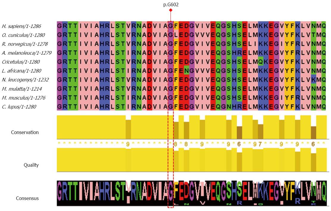
Figure 2 Multiple sequence alignment derived from PolyPhen-2 and edited by Jalview.
Amino acids with similar physicochemical properties have the same color. Aliphatic/hydrophobic residues (I, V, L, A, M) are colored pink, aromatic residues (F, W, Y) orange, positive charged residues (K, R, H) blue, negative charged residues (D, E) red, hydrophilic residues (S, T, N, Q) green, conformationally special (P, G) magenta. I: Isoleucine; V: Valine; L: Leucine; A: Alanine; M: Methionine; F: Phenylalanine; W: Tryptophan; Y: Tyrosine; K: Lysine; R: Arginine; H: Histidine; D: Aspartate; E: Glutamate; S: Serine; T: Threonine; N: Asparagine; Q: Glutamine; P: Proline; G: Glycine.
- Citation: Sun HZ, Shi H, Zhang SC, Shen XZ. Novel mutation in a Chinese patient with progressive familial intrahepatic cholestasis type 3. World J Gastroenterol 2015; 21(2): 699-703
- URL: https://www.wjgnet.com/1007-9327/full/v21/i2/699.htm
- DOI: https://dx.doi.org/10.3748/wjg.v21.i2.699


