Copyright
©The Author(s) 2015.
World J Gastroenterol. Jan 14, 2015; 21(2): 694-698
Published online Jan 14, 2015. doi: 10.3748/wjg.v21.i2.694
Published online Jan 14, 2015. doi: 10.3748/wjg.v21.i2.694
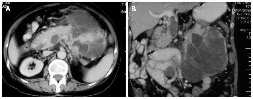
Figure 1 Computed tomography findings.
A: Abdominal computed tomography showing a large heterogenous lesion extending from the body and tail of the pancreas with peripheral enhancement; B: Coronal section showing a thrombus or tumor thrombus in the portal vein and superior mesenteric vein.
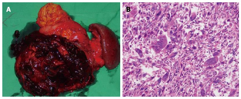
Figure 2 Gross pathology.
A: Gross pathologic examination revealed a 17 cm × 12 cm mass in the pancreatic body and tail. The cut surface of the tumor included a cyst filled with necrotic and hemorrhagic content; B: Microscopically, a mixture of pleomorphic cancer cells and osteoclast-like giant cells were observed with hematoxylin and eosin staining (magnification × 20). Arrows indicate an osteoclast-like giant cell.
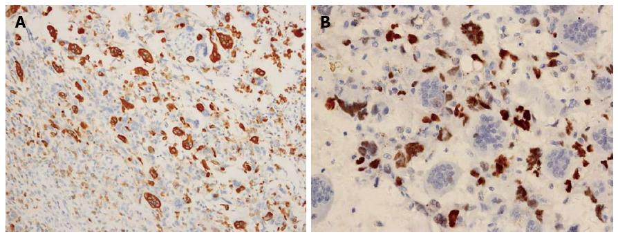
Figure 3 Immunohistochemical findings.
Immunohistochemical assays detected CD68 expression in osteoclast-like giant cells (A) (magnification × 20), and p53 expression in epithelial neoplastic cells (B) (magnification × 40).
- Citation: Gao HQ, Yang YM, Zhuang Y, Liu P. Locally advanced undifferentiated carcinoma with osteoclast-like giant cells of the pancreas. World J Gastroenterol 2015; 21(2): 694-698
- URL: https://www.wjgnet.com/1007-9327/full/v21/i2/694.htm
- DOI: https://dx.doi.org/10.3748/wjg.v21.i2.694


