Copyright
©The Author(s) 2015.
World J Gastroenterol. Jan 14, 2015; 21(2): 688-693
Published online Jan 14, 2015. doi: 10.3748/wjg.v21.i2.688
Published online Jan 14, 2015. doi: 10.3748/wjg.v21.i2.688
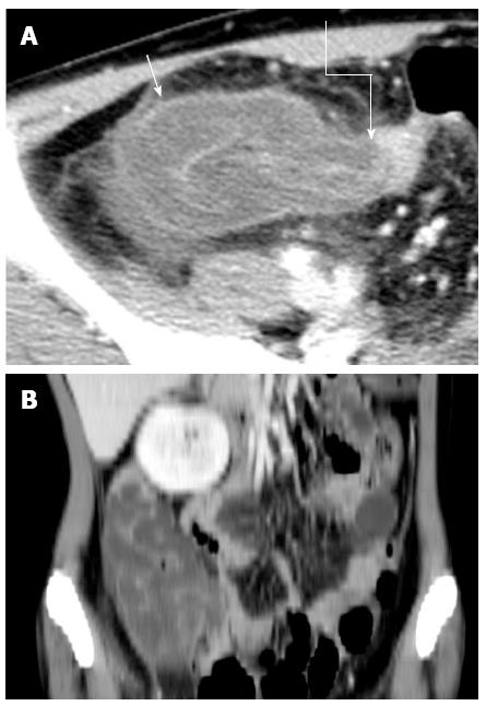
Figure 1 Contrast-enhanced computed tomography image of the lower abdomen.
A: Axial image showed the terminal ileum (intussusceptum, arrow with rugged line) invaginating into the cecum (intussusceptum, arrow with straight line); B: Reformatted oblique coronal image of the iliac fossa showed thickened terminal ileum invaginating into the caecum. The wall of the cecum and ascending colon were thickened and edematous.
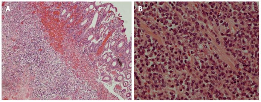
Figure 2 Histological examination of the resected colonic specimen.
A: Low-power examination showed infiltration of leukemic cells into the submucosa; B: High-power examination showed that the leukemia cells were medium-sized with irregular nuclear membranes.
- Citation: Law MF, Wong CK, Pang CY, Chan HN, Lai HK, Ha CY, Ng C, Yeung YM, Yip SF. Rare case of intussusception in an adult with acute myeloid leukemia. World J Gastroenterol 2015; 21(2): 688-693
- URL: https://www.wjgnet.com/1007-9327/full/v21/i2/688.htm
- DOI: https://dx.doi.org/10.3748/wjg.v21.i2.688


