Copyright
©The Author(s) 2015.
World J Gastroenterol. Jan 14, 2015; 21(2): 644-652
Published online Jan 14, 2015. doi: 10.3748/wjg.v21.i2.644
Published online Jan 14, 2015. doi: 10.3748/wjg.v21.i2.644
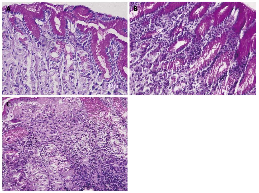
Figure 1 Representative micrographs of gastric corpus mucosal sections showing normal mucosa (A), superficial gastritis (B), and chronic atrophic gastritis (C).
Note the mild infiltration of the gastric mucosa by lymphoid cells near the luminal surface in superficial gastritis (B) and the massive infiltration of the mucosa by lymphoid cells in atrophic gastritis (C).
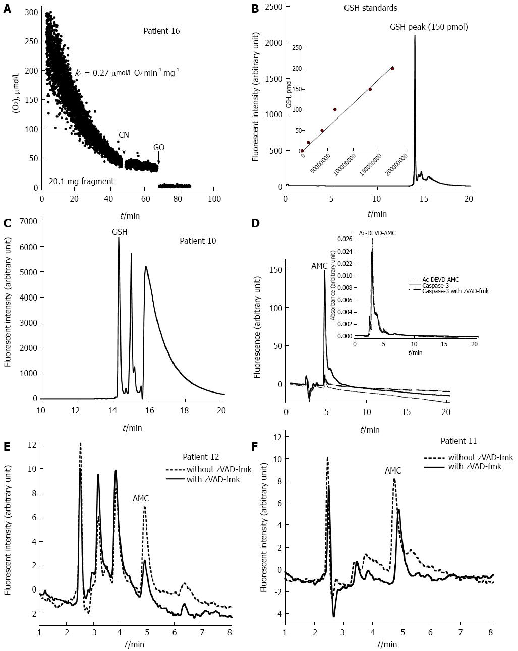
Figure 2 Representative measurements of gastric corpus cellular respiration, glutathione, and caspase activity.
A: A run of cellular mitochondrial O2 consumption by the gastric mucosa of Patient 16. The rate of respiration (k, μmo/L O2 min-1) was set as the negative of the slope of [O2] vs t. The value of kc (μmo/L O2 min-1) and the additions of 10 mmo/L cyanide (a specific inhibitor of cytochrome oxidase) and 50 μg/mL glucose oxidase (catalyzes the reaction of D-glucose + O2 to D-glucono-δ-lactone + H2O2) are shown. O2 consumption was inhibited by cyanide, confirming the oxidation occurred in the mitochondrial respiratory chain. O2 was depleted by the addition of glucose oxidase, confirming the presence of dissolved O2; B: A representative HPLC run of 150 pmol glutathione (GSH) standard (GSH retention time = 14.2 min); GSH standard curve is also shown [insert; GSH (pmol) = 0.00000117 x GSH peak area]; C: A representative HPLC run of cellular GSH in a stomach biopsy (23.9 mg mucosal fragment) from Patient 10. GSH peak area was 354508365 arbitrary units per 50 μL injection volume (reaction volume = 1.0 mL). Thus, cellular GSH content = 347 pmol mg-1 [(354508365 × 0.00000117 × 20)/23.9]; D: Representative HPLC runs for the Ac-DEVD-AMC cleavage reaction by human active caspase-3 with and without the pan-caspase inhibitor zVAD-fmk. The caspase-3 substrate Ac-DEVD-AMC was detected by absorbance at 380 nm with a retention time of about 3 min (insert). The product AMC was detected by fluorescence (380 nm excitation and 460 nm emission) with a retention time of about 4.8 min; E: Representative HPLC runs of caspase activities in the presence (solid line; 22.3 mg mucosal fragment) and absence (dashed line; 21.2 mg mucosal fragment) of the pan-caspase inhibitor zVAD-fmk for Patient 12. The AMC peak area without zVAD-fmk was 16015 arbitrary units/mg and with zVAD-fmk 2810 arbitrary units/mg. Intracellular caspase activity was set as AMC peak area without zVAD-fmk minus with zVAD-fmk, or 13205 (rounded down to 13 × 103) (Table 1); F: Representative HPLC runs of caspase activities with (solid line; 23.3 mg mucosal fragment) and without (dashed line; 19.1 mg mucosal fragment) zVAD-fmk for Patient 11. The AMC peak area without zVAD-fmk was 51207 arbitrary units/mg and with zVAD-fmk 37086 arbitrary unit mg-1. Intracellular caspase activity, thus, was about 14 × 103 (Table 1).
- Citation: Alfazari AS, Al-Dabbagh B, Al-Dhaheri W, Taha MS, Chebli AA, Fontagnier EM, Koutoubi Z, Kochiyi J, Karam SM, Souid AK. Profiling cellular bioenergetics, glutathione levels, and caspase activities in stomach biopsies of patients with upper gastrointestinal symptoms. World J Gastroenterol 2015; 21(2): 644-652
- URL: https://www.wjgnet.com/1007-9327/full/v21/i2/644.htm
- DOI: https://dx.doi.org/10.3748/wjg.v21.i2.644


