Copyright
©The Author(s) 2015.
World J Gastroenterol. Jan 14, 2015; 21(2): 423-431
Published online Jan 14, 2015. doi: 10.3748/wjg.v21.i2.423
Published online Jan 14, 2015. doi: 10.3748/wjg.v21.i2.423
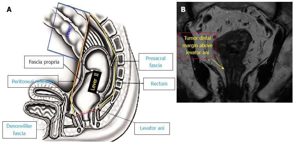
Figure 1 Levels I and II (A), level II (tumor distal margin above levator ani muscle insertion) (B).
Figure 2 Level III (tumor distal margin at the level of levator ani insertion) (A), Level III (B).
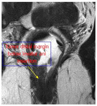
Figure 3 Level IV (tumor distal margin below levator ani insertion).
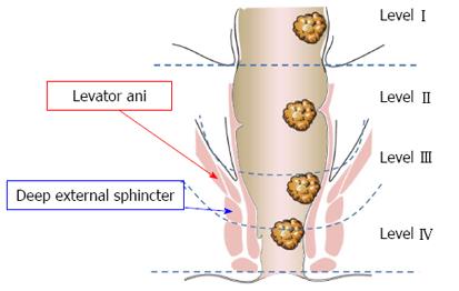
Figure 4 All tumor levels.
- Citation: Alasari S, Lim D, Kim NK. Magnetic resonance imaging based rectal cancer classification: Landmarks and technical standardization. World J Gastroenterol 2015; 21(2): 423-431
- URL: https://www.wjgnet.com/1007-9327/full/v21/i2/423.htm
- DOI: https://dx.doi.org/10.3748/wjg.v21.i2.423


