Copyright
©The Author(s) 2015.
World J Gastroenterol. May 7, 2015; 21(17): 5427-5431
Published online May 7, 2015. doi: 10.3748/wjg.v21.i17.5427
Published online May 7, 2015. doi: 10.3748/wjg.v21.i17.5427
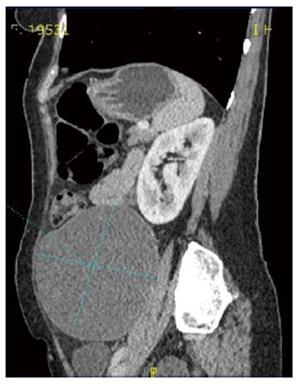
Figure 1 Contrast-enhanced computed tomography scan in the sagittal section showing the retroperitoneal tumor with homogeneous features.
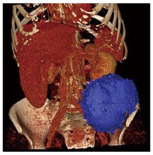
Figure 2 Multidetector computed tomography exam with 3D reconstruction and volumetry.
Imaging revealed an oval, well-delimited, cystic lesion located in the left retroperitoneal space that measured 12.3 cm × 10.8 cm in size (volume: 858 mL) without infiltration of surrounding structures.
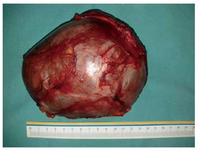
Figure 3 Surgically excised retroperitoneal mass.
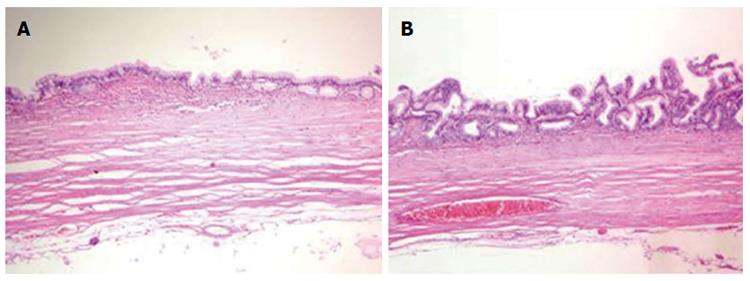
Figure 4 Histopathology.
Hematoxylin eosin staining of the excised tumor revealed a single layer of mucin-producing columnar epithelium with underlying fibrous connective tissue (A, magnification × 14); Rare papillation was observed without invasion (B, magnification × 20).
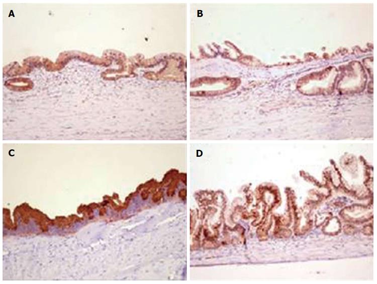
Figure 5 Immunohistochemistry.
Immunoreactive expression of Mucin (MUC) 1 (A); MUC2 (B); MUC5AC (C); and CDX2 (D) (magnification × 20).
- Citation: Knezevic S, Ignjatovic I, Lukic S, Matic S, Dugalic V, Knezevic D, Micev M, Dragasevic S. Primary retroperitoneal mucinous cystadenoma: A case report. World J Gastroenterol 2015; 21(17): 5427-5431
- URL: https://www.wjgnet.com/1007-9327/full/v21/i17/5427.htm
- DOI: https://dx.doi.org/10.3748/wjg.v21.i17.5427


