Copyright
©The Author(s) 2015.
World J Gastroenterol. May 7, 2015; 21(17): 5421-5426
Published online May 7, 2015. doi: 10.3748/wjg.v21.i17.5421
Published online May 7, 2015. doi: 10.3748/wjg.v21.i17.5421
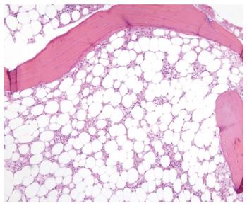
Figure 1 Bone marrow section showing a marked reduction in hemopoietic precursors which are mainly replaced by fat (hematoxylin eosin staining, magnification × 200).
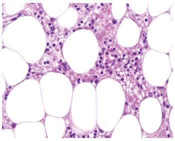
Figure 2 Hematopoietic cells replaced by fat vacuoles and a variable inflammatory infiltrate composed of lymphocytes and plasma cells is observed.
No megakaryocytes are present (hematoxylin eosin staining, magnification × 400).
- Citation: Lens S, Calleja JL, Campillo A, Carrión JA, Broquetas T, Perello C, de la Revilla J, Mariño Z, Londoño MC, Sánchez-Tapias JM, Urbano-Ispizua &, Forns X. Aplastic anemia and severe pancytopenia during treatment with peg-interferon, ribavirin and telaprevir for chronic hepatitis C. World J Gastroenterol 2015; 21(17): 5421-5426
- URL: https://www.wjgnet.com/1007-9327/full/v21/i17/5421.htm
- DOI: https://dx.doi.org/10.3748/wjg.v21.i17.5421


