Copyright
©The Author(s) 2015.
World J Gastroenterol. May 7, 2015; 21(17): 5295-5302
Published online May 7, 2015. doi: 10.3748/wjg.v21.i17.5295
Published online May 7, 2015. doi: 10.3748/wjg.v21.i17.5295
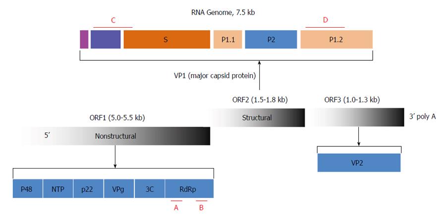
Figure 1 Diagram of the genetic structure of the virus.
The RNA genome is organized into three open reading frames (ORF1, ORF2, and ORF3) that encode the designated nonstructural and structural proteins. The nonstructural protein consists of p48, nucleoside triphosphatase (NTP), p22, viral genome-linked protein (VPg), 3C-like protease (3C), and RdRp. VP1, the major capsid protein, is further organized into N-terminal, shell (S), and protruding (P) domains defined by the indicated VP1 amino acid residues. VP2 is a minor structural protein. The diagnostic and genotyping primers used in the RT-PCR assay targeting conserved areas are shown in red (A-D).
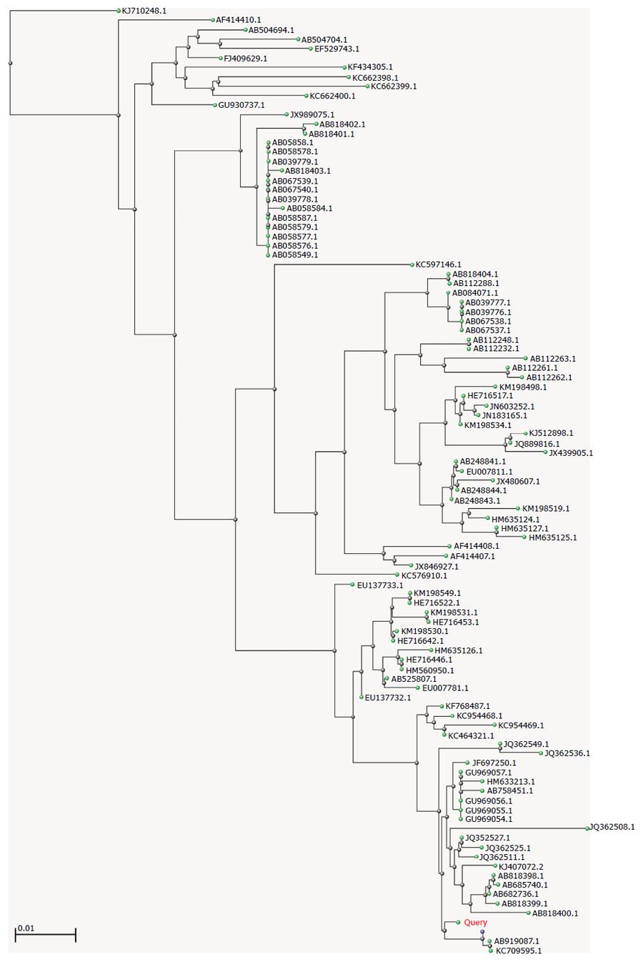
Figure 2 Genetic relatedness among reported genogroup 6 genotype 6 norovirus strains.
The consensus sequence was input into the BLAST, and a distance tree of “100 Blast Hits on the Query Sequence” was drawn using the “Fast Minimum Evolution” method. The accession numbers from the NCBI for all strains are presented. The genetic distance bar is shown in the lower left of the figure.
- Citation: Luo LF, Qiao K, Wang XG, Ding KY, Su HL, Li CZ, Yan HJ. Acute gastroenteritis outbreak caused by a GII.6 norovirus. World J Gastroenterol 2015; 21(17): 5295-5302
- URL: https://www.wjgnet.com/1007-9327/full/v21/i17/5295.htm
- DOI: https://dx.doi.org/10.3748/wjg.v21.i17.5295


