Copyright
©The Author(s) 2015.
World J Gastroenterol. May 7, 2015; 21(17): 5242-5249
Published online May 7, 2015. doi: 10.3748/wjg.v21.i17.5242
Published online May 7, 2015. doi: 10.3748/wjg.v21.i17.5242
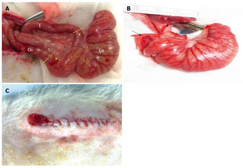
Figure 1 Procedures for intestinal isolation and the creation of an artificial anus on the rat’s dorsum.
A: Anatomical findings reveal the ileum (I), cecum (Ce), and colon (Co). Two cutting points were denoted (dash line) 2-cm apart from ileocecal and cecocolonic junctions (arrowhead); B: The arrow indicates the colonic lumen that remains open; C: The intestinal membrane of the open colonic lumen was sutured together with the rat dorsal skin to create an artificial anus.
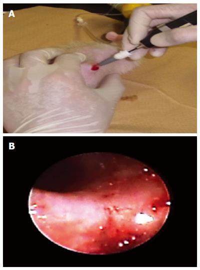
Figure 2 Endoscopic tool used in this animal study.
Figure 3 Internal observation of the cecal pouch using a small animal endoscope via an artificial anus.
A: The endoscope was used to observe the internal cecal pouch via an artificial anus; B: An image of the cecal pouch membrane obtained from the endoscopic examination.
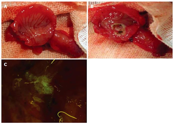
Figure 4 Applications of the newly created isolated cecal pouch model.
A: One-week post-operative surgical re-entry confirmed no fecal contamination of the cecal pouch membrane surface; B: An artificial ulcerative lesion was created on the surface of the pouch membrane; C: Microscopic photograph indicating that cultured small intestinal epithelial cells could be applied to an ulcerative lesion on the cecal pouch membrane.
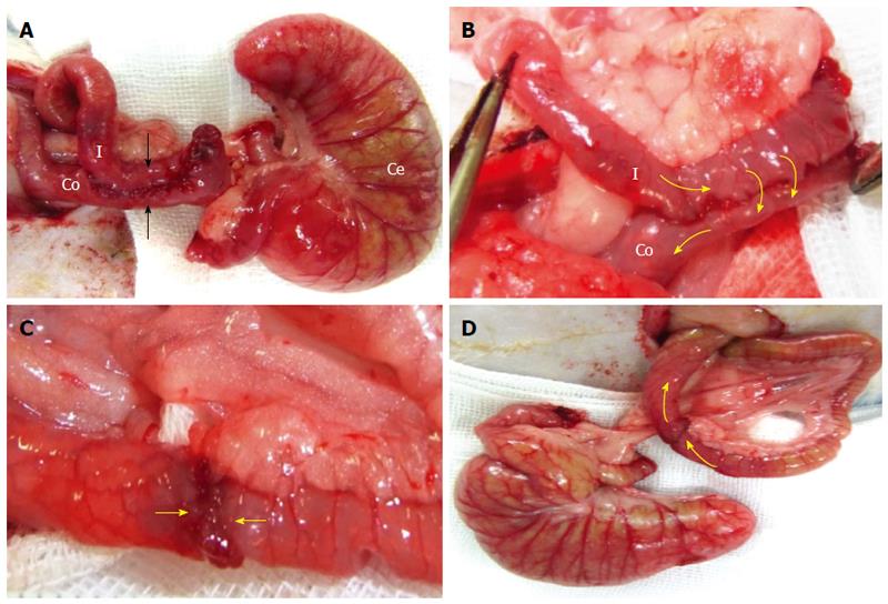
Figure 5 Comparison of two proposed surgical techniques for creating an anastomotic intestine: “side-to-side” anastomosis vs“end-to-end” anastomosis technique.
A: The ileum and colon were anastomosed together using the SSA technique (arrow). An isolated cecal pouch was created; B: The direction of fecal passage from the ileum to the colon via the SSA-anastomotic intestine; C: EEA technique; the frontal surfaces of both cut intestinal lumens were sutured together; D: The direction of fecal passage from the ileum to the colon via the EEA-anastomotic intestine. SSA: “Side-to-side” anastomosis; EEA: “End-to-end” anastomosis.
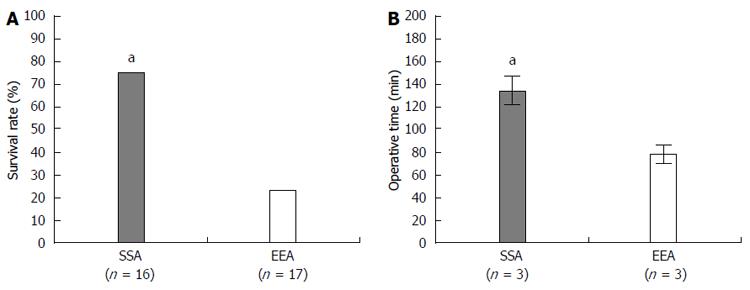
Figure 6 Comparison of the survival rate and operative time between the “side-to-side” anastomosis and end-to-end” anastomosis techniques.
A: Survival rate of animals after creating an anastomotic intestine using the “side-to-side” anastomosis (SSA) or “end-to-end” anastomosis (EEA) technique (aP < 0.05 vs EEA method, Fisher’s exact test); B: Operative time used in the SSA method compared with the EEA method (aP < 0.05 vs EEA method, independent sample-t test).
- Citation: Koshino K, Kanai N, Yamato M, Okano T, Yamamoto M. Novel isolated cecal pouch model for endoscopic observation in rats. World J Gastroenterol 2015; 21(17): 5242-5249
- URL: https://www.wjgnet.com/1007-9327/full/v21/i17/5242.htm
- DOI: https://dx.doi.org/10.3748/wjg.v21.i17.5242


