Copyright
©The Author(s) 2015.
World J Gastroenterol. May 7, 2015; 21(17): 5176-5182
Published online May 7, 2015. doi: 10.3748/wjg.v21.i17.5176
Published online May 7, 2015. doi: 10.3748/wjg.v21.i17.5176
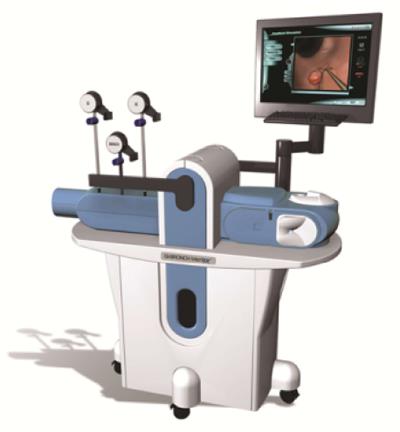
Figure 1 GI-Mentor system.
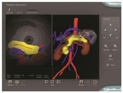
Figure 2 Radial endoscopic ultrasonography image and anatomical 3-dimensional view of pancreas on endoscopic ultrasonography Mentor.
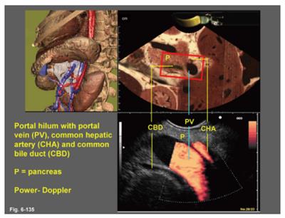
Figure 3 Atlas image of right abdomen on endoscopic ultrasonography Meets VOXEL-MAN (permitted by Dr.
Eike Burmester).
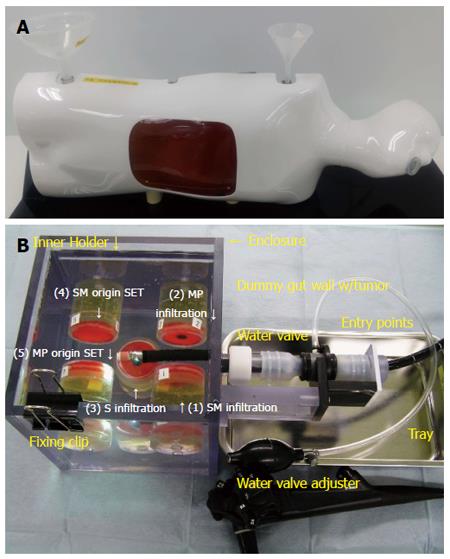
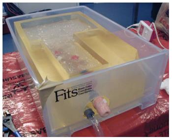
Figure 4 Models of endoscopic ultrasonography phantom.
A: A model for pancreatobiliary system; B: A model for gut wall.
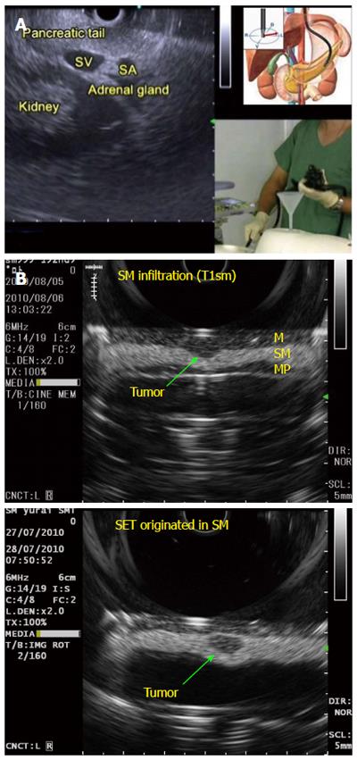
Figure 5 Endoscopic ultrasonography images using endoscopic ultrasonography phantom.
A: Linear endoscopic ultrasonography (EUS) image of pancreas; B: Radial EUS image of submucosal cancer and subepithelial tumor (permitted by Dr. Mitsuhiro Kida).
Figure 6 EUS RK model (permitted by Dr.
Koji Matsuda).
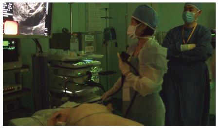
Figure 7 Hands-on-course of endoscopic ultrasonography using a live swine.
- Citation: Kim GH, Bang SJ, Hwang JH. Learning models for endoscopic ultrasonography in gastrointestinal endoscopy. World J Gastroenterol 2015; 21(17): 5176-5182
- URL: https://www.wjgnet.com/1007-9327/full/v21/i17/5176.htm
- DOI: https://dx.doi.org/10.3748/wjg.v21.i17.5176


