Copyright
©The Author(s) 2015.
World J Gastroenterol. May 7, 2015; 21(17): 5119-5130
Published online May 7, 2015. doi: 10.3748/wjg.v21.i17.5119
Published online May 7, 2015. doi: 10.3748/wjg.v21.i17.5119
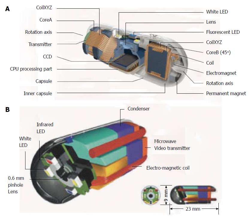
Figure 1 NORIKA and SAYAKA capsules.
A: Sayaka capsule; B: Norika capsule, with permission.
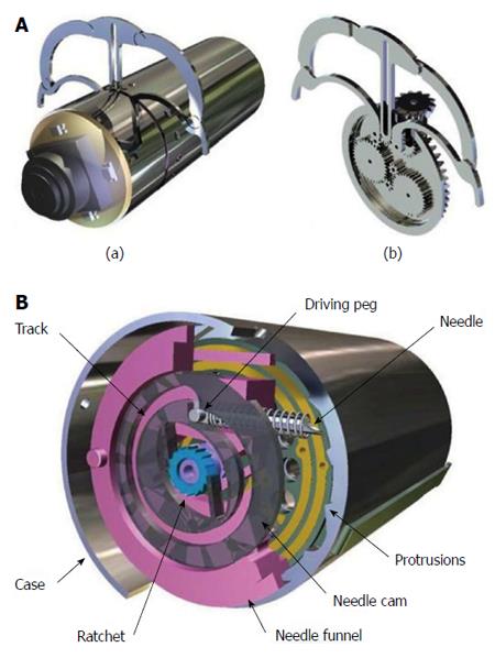
Figure 2 Self-stabilising capsule endoscope.
A: Components; B: Impression of movement while in the colon, with permission.
Figure 3 Odocapsule.
A: Componets; B: Impression of movement inside the bowel, with permission.
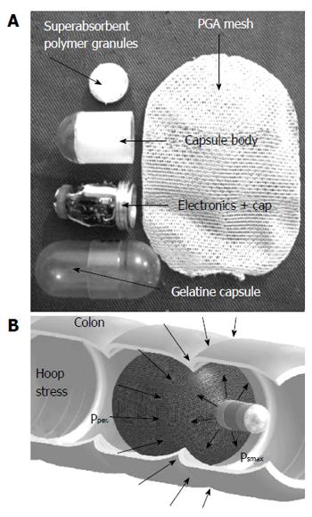
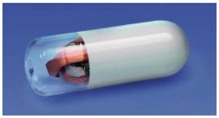
Figure 4 Capsule that provides insufflation, with permission.
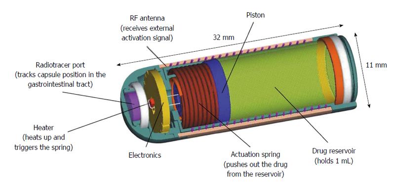
Figure 5 Enterion drug delivery capsule, with permission.
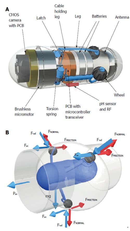
Figure 6 The legged,anchoring capsule for Stabilization of positioning (A) and Drug delivery, with permission (B).
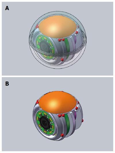
Figure 7 SupCam with (A) and without (B) transparent shell,courtesy of Dr A Tozzi.
Figure 8 Spherical capsule endoscopy with multiple cameras.
Figure 9 3D-Transit" by Motilis Medica SA, Lausanne, Switzerland; courtesy of Mr Vincent Schlageter.
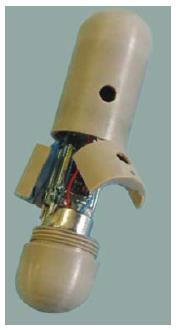
Figure 10 Lab-in-a-Pill, with permission.
- Citation: Koulaouzidis A, Iakovidis DK, Karargyris A, Rondonotti E. Wireless endoscopy in 2020: Will it still be a capsule? World J Gastroenterol 2015; 21(17): 5119-5130
- URL: https://www.wjgnet.com/1007-9327/full/v21/i17/5119.htm
- DOI: https://dx.doi.org/10.3748/wjg.v21.i17.5119


