Copyright
©The Author(s) 2015.
World J Gastroenterol. Apr 28, 2015; 21(16): 5099-5104
Published online Apr 28, 2015. doi: 10.3748/wjg.v21.i16.5099
Published online Apr 28, 2015. doi: 10.3748/wjg.v21.i16.5099
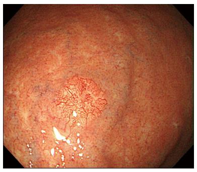
Figure 1 Esophagogastroduodenoscopy findings.
Esophagogastroduodenoscopy revealed a 6 mm, slightly elevated, yellowish lesion with irregular surface vessels at the greater curvature of the upper body.
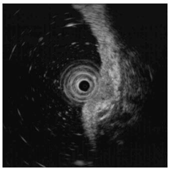
Figure 2 Endoscopic ultrasonography findings.
Endoscopic ultrasonography revealed a 6.7 mm, homogeneous, hypoechoic mass that appeared to invade the submucosal layer.
Figure 3 Endoscopic mucosal resection findings.
A: The inject-lift-and-cut technique was performed; B: Pathologic specimen.
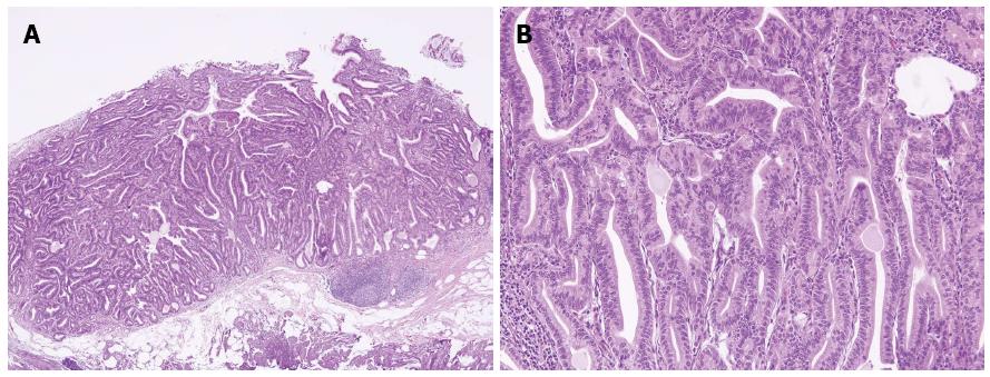
Figure 4 Pathologic examinations.
A: Hematoxylin and eosin staining showed proliferation of glands with complex architecture, situated deep in the mucosal layer with surface involvement (× 40); B: A higher magnification showed complex growth of glands lined with one to two layers of oxyntic epithelium with abundant chief cells. The lining cells showed moderate nuclear pleomorphism, but no mitotic figures were identified (× 200).
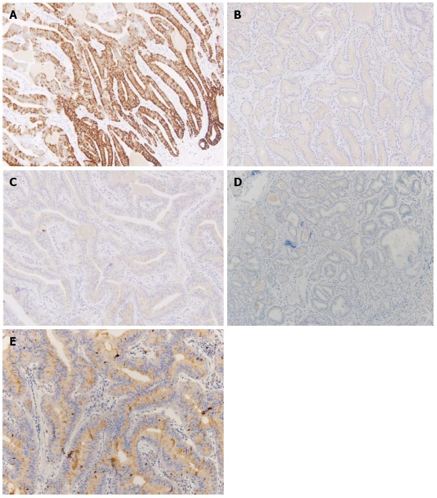
Figure 5 Immunohistochemical staining for mucin, α-fetoprotein, and Ki-67.
A: Most of the glandular cells were positive for mucin (MUC) 6; B: Most of the glandular cells were negative for MUC2; C: Most of the glandular cells were negative for MUC5AC; D: Most of the glandular cells were negative for alpha fetal protein; E: Ki-67 immunolabeling demonstrates a low proliferation index (< 1%).
- Citation: Lee TI, Jang JY, Kim S, Kim JW, Chang YW, Kim YW. Oxyntic gland adenoma endoscopically mimicking a gastric neuroendocrine tumor: A case report. World J Gastroenterol 2015; 21(16): 5099-5104
- URL: https://www.wjgnet.com/1007-9327/full/v21/i16/5099.htm
- DOI: https://dx.doi.org/10.3748/wjg.v21.i16.5099


