Copyright
©The Author(s) 2015.
World J Gastroenterol. Apr 28, 2015; 21(16): 4809-4816
Published online Apr 28, 2015. doi: 10.3748/wjg.v21.i16.4809
Published online Apr 28, 2015. doi: 10.3748/wjg.v21.i16.4809
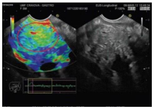
Figure 1 A patient with a malignant pancreatic tumor.
The elastography image in the left panel shows a homogeneous blue mass (red circle). The B-mode reference image is shown in the right panel (Popescu et al[4]).
Figure 2 A patient with chronic pancreatitis.
The elastography image in the left panel shows a heterogeneous green mass (red circle). The B-mode reference image is shown in the right panel (Popescu et al[4]).
Figure 3 Typical contrast-enhanced harmonic endoscopic ultrasound images of pancreatic tumors.
A: Pancreatic carcinoma with hypoenhancement. Conventional EUS (left) shows a hypoechoic mass at the pancreas tail. Contrast-enhanced harmonic endoscopic ultrasound (CH-EUS) (right) indicates that the mass has hypoenhancement compared with the surrounding tissue; B: Chronic pancreatitis with isoenhancement. Conventional EUS (left) shows a hypoechoic mass at the pancreas body. CH-EUS (right) indicates homogeneous enhancement mass similar to the surrounding tissue; a margin is not observed; C: Neuroendocrine tumor with hyperenhancement. Conventional EUS (left) shows a hypoechoic mass at the pancreas body. CH-EUS (right) indicates that enhancement in the mass is higher than in the surrounding tissue (Kwek et al[65]).
- Citation: Meng FS, Zhang ZH, Ji F. New endoscopic ultrasound techniques for digestive tract diseases: A comprehensive review. World J Gastroenterol 2015; 21(16): 4809-4816
- URL: https://www.wjgnet.com/1007-9327/full/v21/i16/4809.htm
- DOI: https://dx.doi.org/10.3748/wjg.v21.i16.4809


