Copyright
©The Author(s) 2015.
World J Gastroenterol. Apr 21, 2015; 21(15): 4583-4591
Published online Apr 21, 2015. doi: 10.3748/wjg.v21.i15.4583
Published online Apr 21, 2015. doi: 10.3748/wjg.v21.i15.4583
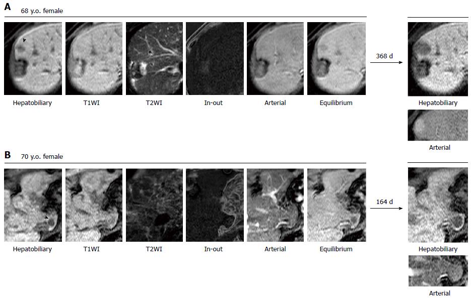
Figure 1 Representative gadolinium ethoxybenzyl diethylene-triamine-pentaacetic-acid magnetic resonance imaging of non-hypervascular nodules that progressed over time.
A: A non-hypervascular nodules (NHNs) approximately 10 mm in diameter was clearly detected in the hepatobiliary phase, as indicated by an arrowhead in the area ventral to the scar of radiofrequency ablation. The NHN was also detected in T1-weighted imaging (T1WI), the arterial phase and the equilibrium phase as a nodule with lower intensity. A fat deposition was unclear in the subtraction of in- and out-of-phase (In-Out) fat-suppressed T1WI. Approximately 1 year later, the NHN had grown to 20 mm in diameter and was partly enhanced in the arterial phase; B: A NHN approximately 8 mm in size was clearly visualized in the hepatobiliary phase, as indicated by an arrowhead in segment 1. The corresponding low-intensity nodule was recognized in T1WI, as well as the equilibrium phase, while a fat deposition was unclear. After approximately 5 mo, the NHN grew to 15 mm in diameter without an increase in the arterial supply.
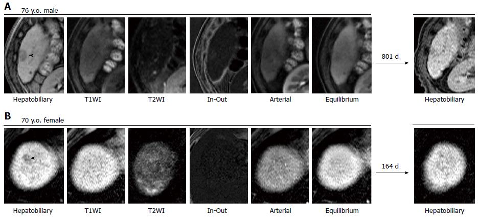
Figure 2 Representative gadolinium ethoxybenzyl diethylene-triamine-pentaacetic-acid magnetic resonance imaging of non-hypervascular nodules that resolved over time.
A: A non-hypervascular nodules (NHNs) was clearly detected at the surface of segment 6 in the hepatobiliary phase, as indicated by an arrowhead in the area ventrolateral to a tiny cyst. The NHN was also revealed in the arterial and equilibrium phases as a lower-intensity nodule, while neither T1-weighted imaging (T1WI) nor T2-weighted imaging visualized the NHN. A fat deposition was expected on the basis of higher intensity in the subtraction of in- and out-of-phase (In-Out) fat-suppressed T1WI. After more than 2 years, the NHN was still there, but the diameter was reduced to less than 5 mm; B: The hepatobiliary phase presented a 6-mm NHN at segment 8 in the vicinity of the diaphragmatic surface of the liver, as indicated by an arrowhead. The NHN could not be visualized in any other sequence. A subsequent gadolinium ethoxybenzyl diethylene-triamine-pentaacetic-acid magnetic resonance imaging showed no nodular lesion in the same area after approximately 5 mo. This NHN was detected together with the other NHN that is presented in Figure 1B.
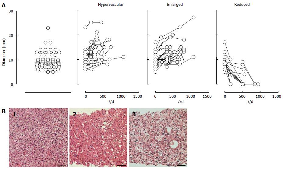
Figure 3 Size distribution over time and representative microscopic images of non-hypervascular nodules that progressed without hypervascularity.
A: The median diameter of 73 non-hypervascular nodules (NHNs) was 9 mm [interquartile range (IQR): 8-12 mm] at the first detection. Diameter was plotted against time to show their growth/reduction rate during the study period. NHNs were classified into 3 groups based on vascularity and size changes in the observation period: hypervascular, enlarged and reduced groups. In the hypervascular group, the vascularity in the arterial phase was confirmed at the endpoint in each case. NHNs in the enlarged group grew at least 2 mm in diameter by the end of the observation, while the diameter decreased 2 mm or more in the reduced group; B: Tissue samples were obtained from 6 of 58 progressed NHNs. Most specimens show thick (1: case #21-1 allocated in supplementary Table 1) or thin (2: case #19-5 allocated in supplementary Table 1) trabecular patterns of tumor cells with nuclear crowding and hypercellularity, which were diagnosed as well-differentiated hepatocellular carcinoma (HCC). One specimen (3: case #22-1 allocated in supplementary Table 1) shows less-differentiated tumor cells with a vacuolated appearance and nuclear irregularity; it was diagnosed as moderately differentiated HCC. (H and E: Each scale bar represents 50 μm.)
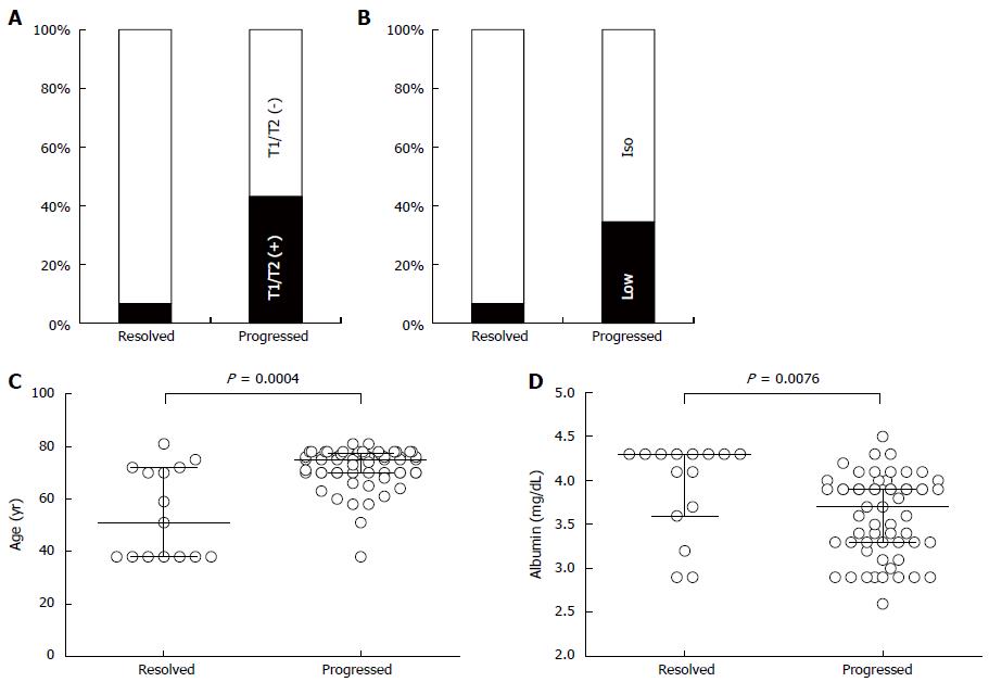
Figure 4 Characteristics of non-hypervascular nodules clarified by univariate analyses.
Non-hypervascular nodules (NHNs) that were detected in the hepatobiliary phase of gadolinium ethoxybenzyl diethylene-triamine-pentaacetic-acid magnetic resonance imaging were univariately characterized. NHNs were classified into 1 of 2 categories, resolved or progressed, in which NHNs decreased by 2 mm or more in diameter without hypervascularity or increased by 2 mm or more or gained an arterial supply, respectively. A: In 58 progressed NHNs, 43% were detected in T1-weighted imaging (T1WI) and/or T2-weighted imaging (T2WI) (black box), whereas only 6.7% showed positive results in the resolved NHNs by T1WI and/or T2WI imaging, leading to a significantly higher detection rate among progressed NHNs; B: In the arterial phase, progressed NHNs were detected as less attenuated nodules (black box) with significantly higher frequency than resolved NHNs were (34.5% vs 6.7%, respectively, P = 0.05); C: The median patient age in the resolved NHN group was 51 years (IQR: 38-72 years), which was significantly younger than the 75 years (IQR: 70-77.3 years) of the progressed group; D: Serum albumin concentration was significantly higher in the resolved NHN group, 4.3 g/dL (IQR: 3.6-4.3 g/dL), than in the progressed group, 3.7 g/dL (IQR: 3.3-3.9 g/dL).
- Citation: Kanefuji T, Takano T, Suda T, Akazawa K, Yokoo T, Kamimura H, Kamimura K, Tsuchiya A, Takamura M, Kawai H, Yamagiwa S, Aoyama H, Nomoto M, Terai S. Factors predicting aggressiveness of non-hypervascular hepatic nodules detected on hepatobiliary phase of gadolinium ethoxybenzyl diethylene-triamine-pentaacetic-acid magnetic resonance imaging. World J Gastroenterol 2015; 21(15): 4583-4591
- URL: https://www.wjgnet.com/1007-9327/full/v21/i15/4583.htm
- DOI: https://dx.doi.org/10.3748/wjg.v21.i15.4583


