Copyright
©The Author(s) 2015.
World J Gastroenterol. Apr 14, 2015; 21(14): 4261-4267
Published online Apr 14, 2015. doi: 10.3748/wjg.v21.i14.4261
Published online Apr 14, 2015. doi: 10.3748/wjg.v21.i14.4261
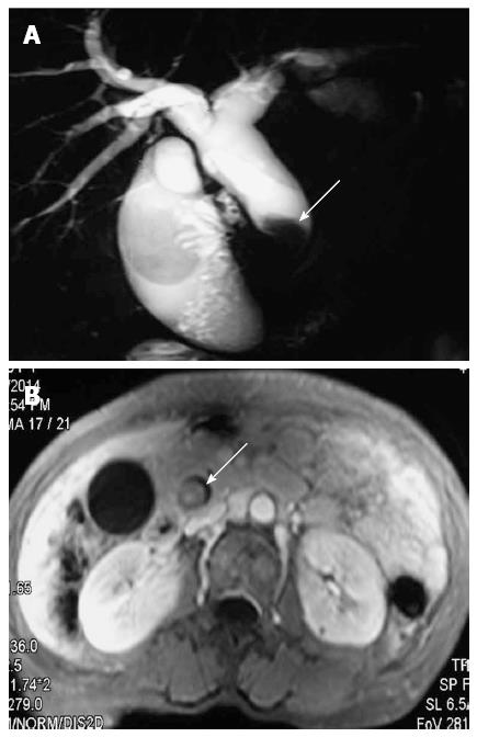
Figure 1 Imaging presentation of biliary tract intraductal papillary mucinous neoplasm.
A: Magnetic resonance cholangiography shows dilation of proximal biliary tract and a filling defect in the extrahepatic biliary tract (arrow); B: Magnetic resonance imaging shows an intraluminal polypoid lesion originating from the extrahepatic biliary tract (arrow).
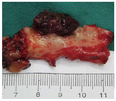
Figure 2 Gross appearance of resected specimen.
Biliary tract intraductal papillary mucinous neoplasm appeared as a nodular lesion on the distal common bile duct with massive mucin deposition throughout.
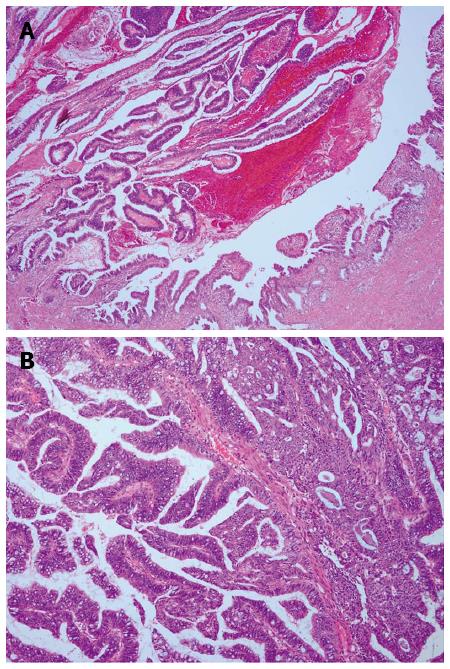
Figure 3 Histopathology of biliary tract intraductal papillary mucinous neoplasm.
Hematoxylin and eosin staining of A: Common bile duct biliary tract intraductal papillary mucinous neoplasm, composed of papillary proliferation of atypical biliary epithelial cells (magnification × 40); and B: High-grade cytologic atypia and mucin in the numerous goblet cells (magnification × 100).
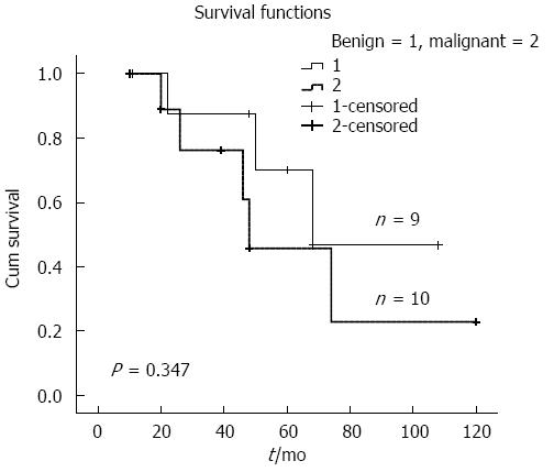
Figure 4 Kaplan-Meier survival curve.
Survival curves for patients with benign (n = 9) and malignant (n = 10) biliary tract intraductal papillary mucinous neoplasms.
- Citation: Wang X, Cai YQ, Chen YH, Liu XB. Biliary tract intraductal papillary mucinous neoplasm: Report of 19 cases. World J Gastroenterol 2015; 21(14): 4261-4267
- URL: https://www.wjgnet.com/1007-9327/full/v21/i14/4261.htm
- DOI: https://dx.doi.org/10.3748/wjg.v21.i14.4261


