Copyright
©The Author(s) 2015.
World J Gastroenterol. Apr 14, 2015; 21(14): 4240-4247
Published online Apr 14, 2015. doi: 10.3748/wjg.v21.i14.4240
Published online Apr 14, 2015. doi: 10.3748/wjg.v21.i14.4240
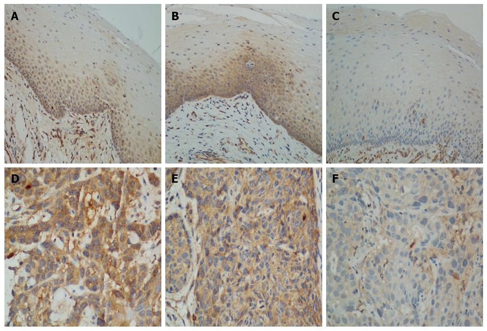
Figure 1 Methyl-methanesulfonate sensitivity 19 immunohistochemistry.
Normal esophageal squamous epithelium with A: Strong nuclear but weak cytoplasmic staining; B: Strong cytoplasmic staining in the basal and suprabasal layers, with scattered strong nuclear staining in the normal epithelium area; C: Weak staining in both the cytoplasm and nucleus (Magnification × 200); Esophageal squamous cell carcinoma with D: Strong staining in the cytoplasm and nucleus; E: Strong staining in the cytoplasm and weak staining in the nucleus; F: Weak staining in both the cytoplasm and nucleus (Magnification × 400).
- Citation: Zhang JL, Wang HY, Yang Q, Lin SY, Luo GY, Zhang R, Xu GL. Methyl-methanesulfonate sensitivity 19 expression is associated with metastasis and chemoradiotherapy response in esophageal cancer. World J Gastroenterol 2015; 21(14): 4240-4247
- URL: https://www.wjgnet.com/1007-9327/full/v21/i14/4240.htm
- DOI: https://dx.doi.org/10.3748/wjg.v21.i14.4240


